AboutKidsHealth


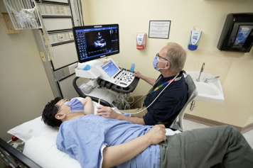
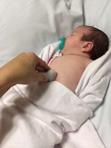
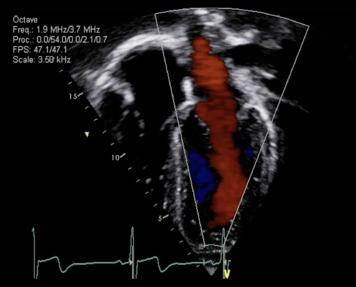
Echocardiogram (heart ultrasound)EEchocardiogram (heart ultrasound)Echocardiogram (heart ultrasound)EnglishCardiologyChild (0-12 years);Teen (13-18 years)HeartHeartTestsCaregivers Adult (19+)NA2021-05-04T04:00:00Z9.4000000000000064.00000000000002022.00000000000Health (A-Z) - ProcedureHealth A-Z<p>An echocardiogram (or ‘echo’) is a special test that uses ultrasound (sound waves) to take pictures of the heart. Learn about how they are done, and how to help your child prepare for one.  </p><h2>What is an echocardiogram?</h2> <figure><span class="asset-image-title">Echocardiogram</span><img alt="Echocardiogram" src="https://assets.aboutkidshealth.ca/AKHAssets/Echocardiogram.png" /> </figure> <p>An echocardiogram (or 'echo' for short) is an ultrasound test of the heart which uses a special ultrasound camera placed on top of the skin and emits sound waves into the body. These sound waves meet structures in the body and reflect (or echo) back to the camera, allowing the health-care team to visualize and assess your child’s heart. </p><h2>Key points</h2><ul><li>An echocardiogram is an ultrasound test of the heart performed by cardiac sonographers or physicians and interpreted by specialized cardiologists. </li><li>Heart rhythm stickers (electrodes), an ultrasound camera and ultrasound jelly over the chest and stomach are used to take pictures of the heart. </li><li>This test lasts between 30 minutes to 90 minutes or more.  </li><li>A child needs to lie still during the test, so some children may need to be sedated or put to sleep for the test. </li></ul><h2>What will happen during the echocardiogram</h2> <figure> <span class="asset-image-title">Echocardiogram room set up</span><img alt="Sonographer doing echocardiogram ultrasound in echo room on patient" src="https://assets.aboutkidshealth.ca/AKHAssets/Echocardiogram%20room%20set%20up.jpg" /><figcaption>The cardiac sonographer uses the ultrasound machine (or echo machine) to take pictures of the heart.</figcaption> </figure> <p>The person who will do the test is specially trained to perform echocardiograms. This person is called a cardiac sonographer. </p><p>The sonographer will first measure your child's weight and height. Then, they will bring you and your child to an echo test room. </p><p>Your child will change from their sweater, shirt and other clothes above the waist and into a gown provided by the sonographer. There are private areas in the echo room to change. </p><p>Your child will lie on a special bed that can move up and down. The sonographer will put three stickers on your child's chest or arms. These stickers are called electrodes. They are connected by wires to the echo machine. They record your child's heartbeat during the test. </p><p>Next, the sonographer will put some warmed ultrasound jelly on your child's chest and belly so that the ultrasound camera can move easily over your child's skin. The sonographer will take pictures of your child's heart from different angles. Your child may feel some gentle pressure from the camera position and movement. Pictures are taken from the stomach, over the chest and from the neck. Your child may also be asked to lie in different positions throughout the test: on their back, on their side, on their back with their head tilted up. At times, your child may hear a loud swooshing noise when the echo machine records the flow of the blood through the heart. </p> <figure> <img alt="Sleeping baby with ultrasound camera on belly" src="https://assets.aboutkidshealth.ca/AKHAssets/Ultrasound%20camera%20on%20infant.jpg" /> <figcaption>The ultrasound camera is placed on the belly of a baby who is comfortably bundled for the echo test. </figcaption> </figure> <p>The lights in the room will be dimmed so the sonographer can see the pictures on the computer screen. </p><p>After the sonographer completes the pictures, they will make a report and show the images to one of the cardiologists, a heart specialist. The cardiologist may choose to take more pictures at this time. This is a normal part of the test. During this time, your child will stay connected to the echo machine. The sonographer or nurse will let you know when your child can get dressed again. </p><p>An echocardiogram takes from 30 minutes to 90 minutes or more. Often the first echocardiogram that your child has will take longer. How long the test lasts will also depend on why your child's doctor asked for the test. </p><h2>Preparing before the echocardiogram</h2><p>Most children do not need to do anything special to get ready for an echocardiogram. However, if your child is under three years old or cannot lie still, they may need a sedative. A sedative is a medicine that will help your child sleep for the test. An echocardiogram works best when the child does not move. </p><h3>Without a sedative</h3><p>If your child does not need a sedative, they can eat and drink normally before and after the echo. If your child has a comforting toy or a security blanket, please bring that along. There is a television in each echo exam room that your child may watch to pass the time. </p><p>Very young babies cannot be given a sedative. This includes babies who are premature or very small. If this is the case for your child, ask the doctor or nurse what the options are. </p><h3>If your child needs a sedative for the echocardiogram</h3><p>Your child needs to lie still during the test so the test results will be accurate. If your child needs a sedative, the bedside nurse will give your child medicine to swallow. This is medicine that will help your child sleep for the test. The medicine lasts just long enough for the test to be done. The nurse will monitor your child throughout the sedation process. </p><p>For sedation, your child must stop eating and drinking several hours before the test. A nurse will call you in the week before the test to make sure you understand the process. The table below describes what and when your child can eat and drink. </p><table class="akh-table"><thead><tr><th>Time before test</th><th>What you need to know</th></tr></thead><tbody><tr><td>8 hours</td><td><p>No more solid food, milk or orange juice. This also means no gum or candy.</p><p>Your child can still drink clear liquids. Clear liquids are anything you can see through, such as apple juice, ginger ale or water.</p><p>Your child can also eat Jell-O or popsicles.</p></td></tr><tr><td>6 hours</td><td>No more milk or formula.</td></tr><tr><td>4 hours</td><td>Stop breastfeeding your baby.</td></tr><tr><td>2 hours</td><td>No more clear liquids. This means no more apple juice, ginger ale or water. Your child cannot eat any Jell-O or popsicles.</td></tr></tbody></table><p>For more information about sedatives, please visit the <a href="/article?contentid=1260&language=english">sedation page</a> or ask your doctor or nurse. </p><h2>General anaesthesia for echocardiograms</h2><p>If your child is older than three years but cannot lie still, they may need <a href="/Article?contentid=1261&language=English">general anaesthesia</a>. Your child will have an appointment in another area of the hospital and there will be an anesthesiologist (sleep doctor) who provides the medication and monitors your child during the procedure. The echo machine will be brought to your child. A nurse will call you the week before the test to make sure you understand how to get ready for the test and the sleep medicine. </p><h2>Role playing the test with your child</h2><p>You and your child can practice the test at home.</p><p>Have your child lie on their back on a bed with the lights dimmed. Tell them that it will be warm and very quiet because the sonographer must concentrate on the study. Tell them the sonographer will put three small stickers on their chest. Put some warm hand lotion on the middle and left side of your child's chest. With the bottom of a smooth drinking glass push the hand lotion around on the chest. Explain that the glass is like the camera that the sonographer uses to take the pictures. Bring along your child's special toy or security blanket. Tell your child they will have to remove their shirt or sweater and wear clothing provided at the echo lab. Tell your child that you may lie down beside them on the echo bed during the test if they wished. Make sure the child knows the test will take some time, but you will never leave them alone. </p><h2>Helping your child prepare for the echocardiogram</h2><p>Be honest and open with your child about what to expect. Explain what will happen during the echocardiogram. Use as much detail as you think is appropriate for your child's level of understanding. Tell them about the appointment in advance so they are not surprised to show up at the hospital. Let your child know that you will be with them during the test.</p><p>The video below can be used to help prepare your child for an echocardiogram, a vascular ultrasound or a bicycle stress echo. </p><div class="asset-video"> <iframe src="https://www.youtube.com/embed/N-Cdm86rwsE?rel=0" frameborder="0"></iframe> </div><h2>At SickKids</h2><p>This test is usually performed in the Echocardiography Laboratory (Echo Lab), part of the <a href="https://www.sickkids.ca/en/care-services/centres/labatt-family-heart-centre/">Labatt Family Heart Centre</a>, located on 4B in the Atrium of the hospital. Sedated echocardiograms are usually performed in the Cardiac Catheterization Lab in the Burton Wing. We also perform bedside echocardiograms for patients throughout the hospital. </p><p>Within the Echo Lab, we also operate the <a href="https://www.sickkids.ca/en/care-services/clinics/cardiology-fetal-echo/">Fetal Cardiac Program</a> as well as our research lab. For more information about our research or if you or someone you know would be interested to participate in cardiovascular research, please visit us at the <a href="https://lab.research.sickkids.ca/cvus/">Cardiovascular Ultrasound Research Lab website</a>. </p>
مخطط صدى القلبممخطط صدى القلبEchocardiogramArabicCardiologyChild (0-12 years);Teen (13-18 years)HeartHeartTestsCaregivers Adult (19+)NA2009-08-19T04:00:00Z000Flat ContentHealth A-Z<p>مخطط صدى القلب هو فحص القلب بالموجات فوق الصوتية. احصل على معلومات عن فحص القلب هذا وما يعني لطفلك إذا كان يحتاج الى مهدّئ لمخطط صدى القلب.</p>
超声心动图(心脏超声波)超声心动图(心脏超声波)Echocardiogram (heart ultrasound)ChineseSimplifiedNAChild (0-12 years);Teen (13-18 years)NANANAAdult (19+)NA2021-05-04T04:00:00Z6.00000000000000Flat ContentHealth A-Z<p>超声心动图(echocardiogram,英文简称“echo”)是利用声波拍摄心脏的照片。了解儿童超声心动图检查的准备工作和具体方法。</p><h2>什么是超声心动图?</h2> <figure> <span class="asset-image-title">超声心动图</span><img alt="超声心动图" src="https://assets.aboutkidshealth.ca/AKHAssets/Echocardiogram.png" /> </figure> <p>超声心动图是一种对心脏的超声波测试,利用一个放置在皮肤上的专用超声波摄像机向体内发射声波。这些声波遇到身体的结构,并反射回(或回声传到)摄像头,使医疗团队能够对孩子的心脏进行可视化和评估。</p><h2>要点</h2><ul><li>超声心动图检查是由心脏超声医师或医生对心脏进行的超声波测试,并由心脏病科医师进行解读。</li><li>心律贴片(电极)、超声波相机以及涂抹在胸部和胃部的超声凝胶用于拍摄心脏照片。</li><li>此检查持续30至90分钟,或更长时间。<br></li><li>在检查过程中,孩子需要躺着不动,所以某些孩子可能需要使用镇静剂或让其睡着。</li></ul>
超聲心動圖(心臟超聲波)超聲心動圖(心臟超聲波)Echocardiogram (heart ultrasound)ChineseTraditionalNAChild (0-12 years);Teen (13-18 years)NANANAAdult (19+)NA2021-05-04T04:00:00ZFlat ContentHealth A-Z<p>超聲心動圖(或「超聲」)是一種運用超聲波(聲波)拍攝心臟圖片的特殊檢測方法。了解檢測如何進行,以及如何協助孩子為檢測作好準備。 </p><h2>什麼是超聲心動圖?</h2> <figure> <span class="asset-image-title">超聲心動圖</span><img alt="超聲心動圖" src="https://assets.aboutkidshealth.ca/AKHAssets/Echocardiogram.png" /> </figure> <p>超聲心動圖(或簡稱「超聲」)是一種心臟超聲檢測方法,運用放置在皮膚表面的特殊超聲攝影器,並將聲波發射到體內。該等聲波在碰到身體結構後會反射(或迴響)回攝影器,讓醫護團隊能觀察和評估孩子的心臟。</p><h2>關鍵重點</h2><ul><li>超聲心動圖是由心臟超聲師或內科醫生對心臟進行的超聲波檢查,並由專門的心臟科醫生解讀。</li><li>拍攝心臟圖片會使用心律貼片(電極),並在胸部與胃部上使用超聲攝影器和超聲波凝膠。</li><li>此檢測需時 30 分鐘至 90 分鐘或以上。 </li><li>檢測期間,孩子需要躺着不動,因此某些孩子或會需要注射鎮靜劑或接受麻醉才能進行檢測。</li></ul>
ÉchocardiogrammeÉÉchocardiogrammeEchocardiogramFrenchCardiologyChild (0-12 years);Teen (13-18 years)HeartHeartTestsCaregivers Adult (19+)NA2009-08-19T04:00:00Z6.0000000000000074.00000000000001308.00000000000Health (A-Z) - ProcedureHealth A-Z<p>Un échocardiogramme utilise des ondes sonores pour prendre des images du cœur. Sachez ce qui se passe pendant un échocardiogramme et comment préparer votre enfant.</p><h2>Qu’est-ce qu’un échocardiogramme?</h2> <figure> <span class="asset-image-title">Échocardiogramme</span><img alt="Échocardiogramme" src="https://assets.aboutkidshealth.ca/akhassets/HC_Echocardiogram_Img_MEDIMG-PHO_EN.jpg" /> </figure> <p>Un échocardiogramme est un examen qui utilise des ondes sonores pour prendre des images du cœur de votre enfant. Un échocardiogramme s’appelle aussi parfois une échocardiographie. Cet examen est sans douleur et entièrement sans danger. Vous pourrez probablement rester avec votre enfant pendant l’examen.</p><h2>Principaux points à retenir</h2> <ul> <li>Un échocardiogramme est un examen qui prend des images du cœur.</li> <li>L’examen ne fait pas mal. Il dure d’une demi-heure à une heure et demie.</li> <li>L’enfant doit rester immobile pendant l’examen; certains enfants devront prendre un sédatif ou seront placés sous anesthésie générale pour la durée de l’examen.</li> <li>Si votre enfant doit prendre un sédatif pour l’examen, il ne doit ni manger ni boire un certain temps avant l’examen. </li> </ul><h2>Que se passe-t-il pendant un échocardiogramme</h2><p>La personne qui fera passer l’examen est un technologue formé tout spécialement pour faire passer des échocardiogrammes. Cette personne est un échographiste.</p><p>L’échographiste commencera par mesurer et par peser votre enfant. Ensuite, il le conduira dans une salle pour les échocardiographies. </p> <p>Votre enfant devra s’allonger sur un lit qui peut se déplacer vers le haut et vers le bas. Il devra enlever son chandail, sa chemise et tout autre vêtement au-dessus de la taille.</p><p>L’échographiste placera trois collants sur la poitrine ou le bras de votre enfant. Ces collants sont des électrodes. Elles sont reliées par des fils à l’appareil pour échocardiogrammes. Elles permettent d’enregistrer les pulsations cardiaques de votre enfant pendant l’examen.</p><p>Ensuite, l’échographiste appliquera un gel sur la poitrine et le ventre de votre enfant pour que la sonde puisse se déplacer facilement sur sa peau. La sonde est un genre de caméra qui prend des photos du cœur de votre enfant. Elle mesure environ 15 centimètres (6 pouces) et son extrémité arrondie repose légèrement sur le gel.</p><p>La plupart des lumières seront éteintes dans la selle pour que l’échographiste puisse voir les images à l’écran de l’ordinateur. L’échographiste déplacera la sonde pour prendre des images du cœur de votre enfant sous divers angles. Il prendra des images depuis l’estomac jusqu’au cou, en passant sur la poitrine. Vous pouvez observer ces images à l’écran de l’ordinateur. Toutes les images sont conservées dans l’ordinateur. </p><p>Votre enfant ne ressentira aucune douleur pendant l’échocardiogramme. Il ressentira une légère pression de la sonde. Parfois, il entendra peut-être un genre de sifflement lorsque l’appareil enregistrera le flux sanguin dans le cœur.</p><p>Une fois que l’échographiste aura terminé de prendre des images, il produira un rapport et montrera les images à un des cardiologues, c’est-à-dire, un spécialiste du cœur. Le cardiologue pourra décider de prendre d’autres images. Cela est normal. Pendant ce temps, votre enfant restera branché à l’appareil pour échocardiogrammes. L’échographiste ou l’infirmière vous indiquera le moment où votre enfant pourra se lever et s’habiller. </p><p>Un échocardiogramme prend de 30 à 90 minutes, ou parfois plus. Souvent, le premier échocardiogramme que subira votre enfant prendra plus de temps. La durée de l’examen dépend des informations que le médecin de votre enfant veut obtenir pendant l’examen. </p><h2>Se préparer à un échocardiogramme</h2> <p>La plupart des enfants n’ont rien à faire pour se préparer à un échocardiogramme. Cependant, si votre enfant a moins de trois ans, il devra peut-être pendre un sédatif. Un sédatif est un médicament qui le fera dormir pendant l’examen. Un échocardiogramme donne de meilleurs résultats lorsque l’enfant ne bouge pas. </p> <h3>Sans sédatif :</h3> <p>Si votre enfant n’a pas besoin de sédatif, il peut manger et boire normalement avant et après l’examen. S’il a un jouet favori, une couverture réconfortante ou un film préféré, apportez-les avec vous pour l’examen.</p> <p>On ne peut pas donner de sédatif aux très jeunes bébés. Cela comprend les bébés prématurés ou très petits. Si c’est le cas de votre enfant, demandez au médecin ou à l’infirmière les options qui s’offrent à vous.</p> <h2>Si votre enfant doit prendre un sédatif pour l’échocardiogramme</h2> <p>Votre enfant devra rester allongé et immobile pendant l’examen pour obtenir de bons résultats. Si votre enfant doit prendre un sédatif, l’infirmière lui donnera un sédatif à avaler. Cela le fera dormir pendant la durée de l’examen, pas plus. </p> <p>Si vous voulez en savoir plus au sujet des sédatifs, adressez-vous au médecin ou à l’infirmière.</p> <h3>Manger ou boire avant un échocardiogramme</h3> <p>Si votre enfant doit prendre un sédatif pour l’examen, il doit cesser de manger et de boire plusieurs heures avant l’examen. Une infirmière vous appellera au cours de la semaine qui précède l’examen pour s’assurer que vous comprenez bien ce que votre enfant peut, et ne peut pas, manger et boire. Le tableau ci-dessous indique le moment où votre enfant doit cesser de manger et de boire. </p> <h3>Ce que votre enfant peut manger ou boire avant le sédatif</h3> <table class="akh-table"> <thead> <tr><th>Temps avant l’examen</th><th>Ce qu’il faut savoir</th></tr> </thead> <tbody> <tr> <td>8 heures</td> <td><p>Plus aucune nourriture solide, lait ou jus d’orange. Cela signifie également aucune gomme à mâcher ou aucuns bonbons.</p><p>Votre enfant peut encore boire des liquides clairs. Les liquides clairs sont ceux qui sont translucides (on peut voir à travers) comme le jus de pomme, le ginger ale ou l’eau.</p><p>Votre enfant peut aussi manger du Jell-O ou des jus glacés (popsicles).</p></td> </tr> <tr> <td>6 heures</td> <td>Plus de lait ou de formules lactées.</td> </tr> <tr> <td>4 heures</td> <td>Arrêtez d’allaiter votre bébé.</td> </tr> <tr> <td>2 heures</td> <td>Plus de liquides clairs. Cela signifie plus de jus de pomme, d’eau ou de ginger ale, de Jell-O et de jus glacés (popsicles).</td> </tr> </tbody> </table> <h2>Anesthésie générale pour les échocardiogrammes</h2> <p>Si votre enfant a plus de trois ans, mais qu’il ne peut rester allongé et immobile, il aura peut-être besoin d’une anesthésie générale (sédatif). Il s’agit d’un médicament pour dormir administré par un médecin. Pour cela, votre enfant recevra un rendez-vous dans un autre secteur de l’hôpital. L’échocardiographe (appareil pour les échocardiogrammes) sera apporté là où se trouvera votre enfant. Une infirmière vous appellera la semaine avant l’examen pour s’assurer que vous savez comment vous préparer à l’examen et au médicament pour dormir. </p> <h2>Aider votre enfant à se préparer à l’échocardiogramme</h2> <p>Soyez honnête et parlez ouvertement à votre enfant; dites-lui ce à quoi il doit s’attendre et ce qui se passera pendant l’échocardiogramme. Utilisez autant de détails que vous jugez appropriés pour que votre enfant comprenne. Informez-le du moment où il aura son rendez-vous afin qu’il ne soit pas surpris de devoir se rendre à l’hôpital. Dites-lui que l’examen ne fait pas mal et qu’il n’a pas à avoir peur. Dites-lui que vous l’accompagnerez pendant l’examen.</p> <h2>Imitez le déroulement de l’examen avec votre enfant</h2> <p>Si vous le voulez, avec votre enfant vous pouvez pratiquer le déroulement de l’examen à la maison. Demandez-lui de s’allonger sur le dos dans une salle obscure. Dites-lui qu’il fera chaud et qu’il n’y aura pas de bruit, car l’échographiste doit se concentrer.</p> <p>Appliquez de la crème hydratante depuis le centre de sa poitrine sur tout le côté gauche. À l’aide d’un verre à fond plat et lisse, badigeonnez la crème sur sa poitrine. Expliquez à votre enfant que le verre imite la caméra que l’échographiste utilisera pour prendre des images.</p> <p>Apportez un jouet, une couverture ou un film que votre enfant aime.</p> <p>Dites à votre enfant que pour l’examen, il devra retirer sa chemise ou son chandail et il devra porter les vêtements qui lui seront remis au laboratoire.</p> <p>Dites-lui que vous vous allongerez à côté de lui sur le lit pendant l’examen. Assurez-vous qu’il comprend que l’examen prendra du temps, mais que vous ne le laisserez pas seul.</p><h2>À l’hôpital SickKids :</h2> <p>Cet examen est habituellement pratiqué dans le secteur 4B de l’atrium de l’hôpital. Chacune des salles comprend un magnétoscope et une télévision que votre enfant peut regarder pendant l’examen.</p>
EcocardiogramaEEcocardiogramaEchocardiogramPortugueseNAChild (0-12 years);Teen (13-18 years)NANANAAdult (19+)NA2009-08-19T04:00:00Z74.00000000000006.000000000000001308.00000000000Flat ContentHealth A-Z<p>Ecocardiograma infantil é uma exame ao coração da criança. Leia e aprenda tudo sobre a preparação para um ecocardiograma para a sua criança.</p>
ਐਕੋਕਾਰਡੀਓਗਰਾਮਐਕੋਕਾਰਡੀਓਗਰਾਮEchocardiogramPunjabiNAChild (0-12 years);Teen (13-18 years)NANANAAdult (19+)NA2009-08-19T04:00:00Z000Flat ContentHealth A-Z<p>ਐਕੋਕਾਰਡੀਓਗਰਾਮ ਦੌਰਾਨ ਕੀ ਕੁਝ ਹੁੰਦਾ ਅਤੇ ਆਪਣੇ ਬੱਚੇ ਨੂੰ ਇਸ ਲਈ ਕਿਵੇਂ ਤਿਆਰ ਕਰਨਾ ਹੈ ਬਾਰੇ ਸਿਖਿਆ ਹਾਸਲ ਕਰੋ।</p>
EcocardiogramaEEcocardiogramaEchocardiogramSpanishNAChild (0-12 years);Teen (13-18 years)NANANAAdult (19+)NA2009-08-19T04:00:00Z000Flat ContentHealth A-Z<p>El resultado de un ecocardiograma son imágenes del corazón tomadas por ondas sonoras. Infórmese sobre cómo preparar a su niño para hacerse un ecocardiograma.</p>
மின் ஒலி இதய வரைவு (எக்கோ கார்டியோகிராம்)மின் ஒலி இதய வரைவு (எக்கோ கார்டியோகிராம்)EchocardiogramTamilNAChild (0-12 years);Teen (13-18 years)NANANAAdult (19+)NA2009-08-19T04:00:00Z000Flat ContentHealth A-Z<p>மின் ஒலி இதய வரைவானது இதயத்தின் படங்களை எடுக்க ஒலி அலைகளை உபயோகப்படுத்துகிறது.</p>
ایکو کارڈیو گراماایکو کارڈیو گرامEchocardiogramUrduNAChild (0-12 years);Teen (13-18 years)NANANAAdult (19+)NA2009-08-19T04:00:00Z74.00000000000006.000000000000001308.00000000000Flat ContentHealth A-Z<p>ایکو ڈائیگرام میں دل کی تصویر لینے کیلئے صوتی لہروں کا استعمال کیا جاتا ہے۔ اس طریق کار اور اپنے بچے کو ایکو ڈائیگرام ٹیسٹ کیلئے تیار کرنے کے طریقے کے بارے میں مزید پڑھیں۔</p><br>


超聲心動圖(心臟超聲波)1274.00000000000超聲心動圖(心臟超聲波)Echocardiogram (heart ultrasound)ChineseTraditionalNAChild (0-12 years);Teen (13-18 years)NANANAAdult (19+)NA2021-05-04T04:00:00ZFlat ContentHealth A-Z<p>超聲心動圖(或「超聲」)是一種運用超聲波(聲波)拍攝心臟圖片的特殊檢測方法。了解檢測如何進行,以及如何協助孩子為檢測作好準備。 </p><h2>什麼是超聲心動圖?</h2> <figure> <span class="asset-image-title">超聲心動圖</span><img alt="超聲心動圖" src="https://assets.aboutkidshealth.ca/AKHAssets/Echocardiogram.png" /> </figure> <p>超聲心動圖(或簡稱「超聲」)是一種心臟超聲檢測方法,運用放置在皮膚表面的特殊超聲攝影器,並將聲波發射到體內。該等聲波在碰到身體結構後會反射(或迴響)回攝影器,讓醫護團隊能觀察和評估孩子的心臟。</p><h2>關鍵重點</h2><ul><li>超聲心動圖是由心臟超聲師或內科醫生對心臟進行的超聲波檢查,並由專門的心臟科醫生解讀。</li><li>拍攝心臟圖片會使用心律貼片(電極),並在胸部與胃部上使用超聲攝影器和超聲波凝膠。</li><li>此檢測需時 30 分鐘至 90 分鐘或以上。 </li><li>檢測期間,孩子需要躺着不動,因此某些孩子或會需要注射鎮靜劑或接受麻醉才能進行檢測。</li></ul><h2>超聲心動圖檢測前的準備</h2><p>大多數孩子毋需為接受超聲心動圖檢測而作特別的準備。可是,如果孩子不足 3 歲或不能靜臥,則他們或會需要使用鎮靜劑。鎮靜劑是一種藥物,有助孩子在睡眠中完成檢測。當孩子身體不動時,超聲心動圖的效果最佳。</p><h3>無需鎮靜劑</h3><p>如果孩子不需要鎮靜劑,則可在超聲檢測前後正常飲食。如果孩子有喜愛的玩具或安撫毯,請隨身攜帶。每間超聲檢測室都配備一台電視,孩子可以觀看電視以打發時間。</p><p>太小的嬰兒不能使用鎮靜劑,其中包括早產或體重較輕的嬰兒。如果您的孩子屬於此類情況,請諮詢醫生或護士,以了解合適的選擇。</p><h3>如果孩子需要鎮靜劑以進行超聲心動圖檢測</h3><p>孩子需要在檢測期間保持靜臥,以確保檢測結果準確。如果孩子需要使用鎮靜劑,則臨床護士會給孩子服藥。這種藥物有助孩子在睡眠中完成檢測。藥效持續時間僅足以完成檢測。護士會在鎮靜期間全程監察孩子。</p><p>如要使用鎮靜劑,則孩子必須在檢測前數小時停止飲食。護士會在檢測前一週來電,以確保您了解整個檢測過程。下表描述了孩子何時可以進食和飲用什麼。</p><table class="akh-table"><thead><tr><th>檢測前</th><th>須知</th></tr></thead><tbody><tr><td>8 小時</td><td><p>不再進食固體食物,或飲用牛奶或橙汁。這亦意味着不能食用口香糖或糖果。</p><p>孩子可飲用清流質。清流質是可透視的清澈液體,如:蘋果汁、薑汁汽水或水。</p><p>孩子亦可以食用果凍或冰棒。</p></td></tr><tr><td>6 小時</td><td>不再飲用牛奶或配方奶。</td></tr><tr><td>4 小時</td><td>停止母乳餵哺嬰兒。</td></tr><tr><td>2 小時</td><td>不再飲用清流質。這意味着不可以再飲用蘋果汁、薑汁汽水或水。孩子亦不能食用果凍或冰棒。</td></tr></tbody></table><p>有關鎮靜劑的詳情,請瀏覽鎮靜頁面,或諮詢醫生或護士。</p><h2>全身麻醉以進行超聲心動圖檢測</h2><p>如果孩子大於 3 歲但不能靜臥,則或會需要 <a href="/article?contentid=1261&language=chinesetraditional">全身麻醉</a>。孩子會在醫院的另一個區域就診,在該處麻醉師會施用麻醉藥,並在麻醉期間全程監察孩子。超聲檢測儀會被帶到孩子接受麻醉的地方。護士會在檢測前一週來電,以確保您了解如何為檢測和麻醉作好準備。</p><h2>透過角色扮演與孩子模擬檢測過程</h2><p>您和孩子可在家中模擬檢測過程。</p><p>將燈光調暗,讓孩子仰臥。告訴他們室內會很溫暖且非常安靜,因為超聲師必須全神貫注地進行檢測。告訴孩子,超聲師會在其胸部貼上三個小貼片。在孩子胸部正中和左側塗上一些溫熱的護手霜。用光滑的玻璃杯底部在胸部推開護手霜,並解釋說玻璃杯就像超聲師用來拍照的攝影器。帶上孩子最愛的玩具或安撫毯。告訴孩子必須脫掉襯衫或毛衣,然後穿上超聲實驗室提供的衣服。告訴孩子,您可以按他們意願,在檢測期間躺在超聲床上,躺在他們身旁。確保孩子知道檢測需要一段時間,但您會一直陪伴。</p><h2>協助孩子為超聲心動圖檢測作好準備</h2><p>誠實且坦率地告訴孩子將會發生什麼。耐心解釋超聲心動圖檢測的過程是怎樣。以切合孩子理解水平的方式,儘量詳細解釋。提前告訴他們檢測時間,這樣他們便不會對要到醫院感到驚訝。告訴孩子,您會在檢測期間一直陪伴。</p><p>以下影片可用於協助孩子為超聲心動圖、血管超聲波或單車負荷超聲檢測作好準備。</p><div class="asset-video"> <iframe src="https://www.youtube.com/embed/N-Cdm86rwsE?rel=0" frameborder="0"></iframe> </div><h2>超聲心動圖檢測的過程</h2> <figure><span class="asset-image-title">超聲心動圖室的佈置</span> <img src="https://assets.aboutkidshealth.ca/AKHAssets/Echocardiogram%20room%20set%20up.jpg" alt="" /> <figcaption>心臟超聲師運用超聲檢測儀(或迴聲器)拍攝心臟圖片。</figcaption> </figure> <p>檢測師受過進行超聲心動圖檢查的專門培訓。他們被稱為心臟超聲師。</p><p>超聲師會首先量度孩子的身高和體重。隨後,他們會將您和孩子帶到超聲檢測室。</p><p>孩子要將腰部以上的毛衣、襯衫和其他衣物更換為超聲師所提供的長袍。超聲室設有私人的更衣區。</p><p>孩子會躺在一張可上下移動的床上。超聲師會在孩子的胸部或手臂貼上三個貼片。這些貼片稱為電極,由電線連接至超聲檢測儀,並在檢測期間記錄孩子的心跳。</p><p>接下來,超聲師會在孩子的胸部和腹部塗上一些溫熱的超聲波凝膠,以便超聲波攝影器能輕易地在孩子的皮膚上移動。超聲師會從不同角度拍攝孩子的心臟照片。孩子或會在攝影器的所在位置和移動期間感到輕微的按壓感。攝影器會從胃部、胸部和頸部拍攝照片。在檢測過程中,或會要求孩子以不同的姿勢躺着:仰臥、側臥、仰臥時仰頭。有時,當超聲檢測儀記錄心臟的血流時,孩子或會聽到響亮的嗖嗖聲。</p> <figure> <img src="https://assets.aboutkidshealth.ca/AKHAssets/Ultrasound%20camera%20on%20infant.jpg" alt="" /> <figcaption>超聲波攝影器會放置在嬰兒的腹部,而嬰兒會在舒適地被捆綁的狀態下進行超聲檢測。</figcaption> </figure> <p>室內燈光會調暗,以便超聲師能看清電腦屏幕上的圖片。</p><p>超聲師會在完成拍攝後製作報告,並向心臟科醫生展示圖片。此時,心臟科醫生或會選擇拍攝更多照片。這是檢測的正常步驟。在此期間,孩子會與超聲檢測儀保持連接。超聲師或護士會通知您孩子何時可以換回自己的衣服。</p><p>超聲心動圖檢測需時 30 分鐘至 90 分鐘或以上。通常,孩子第一次做超聲心動圖檢測會需時較久。檢測所需的時間亦取決於醫生要求孩子進行檢測的原因。</p><h2>其他類型的超聲心動圖</h2><h3>什麼是胎兒超聲心動圖?</h3><p>當嬰兒仍在母親子宮內時所進行的超聲心動圖檢測,稱為胎兒超聲心動圖。超聲心動圖檢測可以在懷孕 16 週時檢測到畸胎,此時胎兒的心臟已清晰可見。如有先天性心臟病家族史、懷疑染色體異常、心率不整,或母親患有可能會影響胎兒心臟發育的疾病,則通常會進行胎兒超聲心動圖檢測。</p><p>在進行此檢測時,會在孕婦胃部塗上超聲波凝膠,這有助傳送聲波並獲取高質素的照片。進行此檢測前通常不需喝水。</p><p>詳情請瀏覽 <a href="https://www.sickkids.ca/en/care-services/clinics/cardiology-fetal-echo/">胎兒心臟計劃網站</a>。</p><h3>什麼是運動負荷超聲心動圖? </h3><p>此超聲心動圖檢測是在孩子騎行一種特殊的單車時進行。心臟圖片會在孩子開始騎單車前、騎行中,以及運動後採集。在整個檢測過程中,亦會監測孩子的血壓和心電活動。</p><p>這項檢測用於研究在非靜止狀態下,心肌收縮以及血液流經心瓣和冠狀動脈的情況。此檢測通常用於了解運動對心臟的影響,以及運動後心臟恢復的速度。</p><h3>什麼是多巴酚丁胺負荷超聲心動圖? </h3><p>多巴酚丁胺負荷超聲心動圖是一種負荷超聲心動圖檢測的形式,在經靜脈注射名為多巴酚丁胺的藥物(含或不含鎮靜劑)後進行。多巴酚丁胺是一種可加速心臟搏動的藥物,其對心臟的作用與運動相似。多巴酚丁胺不會長時間留在體內,亦沒有持久的影響。如果不使用鎮靜劑,孩子在檢測期間或會感到焦慮,因為他們會感到心跳像在運動時般加快,且其感知溫度亦或會上升。進行此檢測方法的原因與運動負荷超聲心動圖相似,但可用於無法進行運動以接受檢測的兒童。</p><h3>什麼是氣泡測試? </h3><p>如有需要,在進行超聲心動圖檢測期間,會將一種稱為造影劑的液體注入孩子的血液中。這有助獲取更清晰的超聲心動圖影像。其中一種造影劑是混合氣體的鹽水(無菌鹽水)。通常採用二氧化碳氣體,亦即令梳打水起泡的同一種氣體。若運用此檢測方法,即稱為氣泡測試。</p><p>氣泡測試可讓心臟科醫生觀察氣泡通過血流的路徑。這有助發現心臟或肺部問題。氣泡測試對人體無害,且氣泡溶液很容易被孩子的血液吸收。 </p><h3>什麼是經食道超聲心動圖 (TEE)?</h3><p>經食道超聲心動圖 (TEE) 是透過將特殊探頭放入喉嚨並伸入食道所進行的超聲心動圖檢測。探頭是一根幼長軟管,末端帶有一個微型攝影器。檢測期間,孩子會接受全身麻醉。</p><p>經食道超聲心動圖可拍攝比常規超聲更清晰的圖片,因為探頭能更接近心臟。如果常規超聲心動圖的圖片不夠清晰,則會在手術結束前,在手術室內,以及有時在心導管插入術期間進行 TEE。</p><h2>獲取超聲心動圖檢測結果</h2><p>只有孩子的心臟科醫生、普通科醫生或其他要求檢測的專科醫生,才能為您提供孩子的超聲心動圖檢測結果。</p><p>進行檢測的超聲師或審閱圖片的心臟科醫生皆不得向您提供檢測結果。這一點很重要,因為他們並不了解您和您孩子的所有健康狀況。</p><h2>在 SickKids</h2><p>此檢測通常在醫院 Atrium 4B 的 <a href="https://www.sickkids.ca/en/care-services/centres/labatt-family-heart-centre/">Labatt 家庭心臟中心 (Labatt Family Heart Centre)</a> 的超聲心動圖實驗室 (超聲室) 進行。使用鎮靜劑的超聲心動圖檢測通常在 Burton Wing 的心導管實驗室 (Cardiac Catheterization Lab) 進行。我們亦為全院病人提供床邊超聲心動圖檢測服務。</p><p>超聲室內亦設有 <a href="https://www.sickkids.ca/en/care-services/clinics/cardiology-fetal-echo/">胎兒心臟計劃</a>以及研究實驗室。如欲了解更多的研究資訊,或者如果您或您認識的人有興趣參與心血管研究,請瀏覽 <a href="https://lab.research.sickkids.ca/cvus/">心血管超聲研究實驗室 (Cardiovascular Ultrasound Research Lab)</a> 網站。</p>https://assets.aboutkidshealth.ca/AKHAssets/Echocardiogram.png超聲心動圖(心臟超聲波)False

Thank you to our sponsors

AboutKidsHealth is proud to partner with the following sponsors as they support our mission to improve the health and wellbeing of children in Canada and around the world by making accessible health care information available via the internet.

Our Sponsors