The brain is an organ located inside your head. The brain and the spinal cord form the central nervous system. This complex system controls everything we do. Each part of the brain has a different job.
The following are some examples of functions that the brain controls:
- Movement, such as walking or stretching
- The 5 senses (seeing, smelling, touching, tasting, and hearing)
- Emotions, thoughts and memory
- Breathing and heartbeat
- Digesting food
- Talking and understanding what others say
The brain is like a busy city. Each part has different functions and is made up of different types of cells. To work, different parts of the brain need to send messages to each other, and to other parts of the body.
What is the brain made of?
Neurons and neuroglial cells
The brain is made up of two types of cells. One type is nerve cells, which are called neurons. A neuron is a nerve cell that sends and receives messages.
The other type is supporting cells, which are called neuroglial cells. Neuroglial cells, or neuroglia, protect and support nerve cells (neurons). These cells are also called glia or glial cells. Some different types of neuroglial cells are oligodendroglia, astrocytes and ependymal cells.
There about 50 neuroglial cells for each neuron cell found in the brain.
The cortex, grey matter and white matter
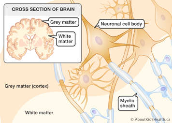
The surface of the cerebrum, called the cortex, is made up of the cell bodies of neurons and neuroglial cells. Because of its colour, the cortex is called “grey matter ." Underneath the cortex, the neuroglial cells and the axons of the neurons form “white matter.” Axons are like wires that carry messages between neurons.

The meninges
The meninges are three thin layers of tissue that protect the brain and spinal cord from injury. The layers are called the dura mater, arachnoid and pia mater. Cerebrospinal fluid flows in between the arachnoid and pia mater membranes. This area is called the subarachnoid space.
This is a supplemental animation that shows cerebrospinal fluid (CSF) moving through the ventricles of the brain. The text in this animation repeats information from the paragraphs above.
Cerebrospinal fluid (CSF)
CSF is a clear fluid that is produced by cells in the ventricles (fluid-filled spaces in the brain). CSF flows through the ventricles and in the space between the layers of tissue that cover the brain and spinal cord (meninges). CSF carries nutrients from the blood to the brain and spinal cord, removes waste products from the brain and cushions and supports the brain and spinal cord.
The cranium
The brain is covered by bones called the cranium. The cranium, together with other bones that protect the face, form the skull.
What is the spinal cord made of?
The spinal cord is made up of neurons that connect the brain to most parts of the body. The spinal cord is covered by the meninges. Like the brain, the spinal cord is cushioned by CSF. The spinal cord is protected by a bony covering called the vertebral or spinal column.
Parts of the brain
There are three major parts of the brain: the cerebrum, the cerebellum and the brainstem.
What does the cerebrum do?

The cerebrum is often used as another word for the brain. It is the largest part of the brain and fills most of the upper skull. The cerebrum uses information from our 5 senses to help us understand what is happening around us. Then it tells our body how to respond. It also controls our emotions, and our ability to talk, think, read, and learn. The surface of the cerebrum is called the cerebral cortex or "grey matter." Underneath the surface is the "white matter."
The cerebrum has two parts: the right and left cerebral hemispheres. In most people, they perform the following functions:
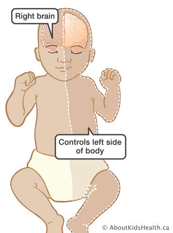
- The left hemisphere plays an important role in language, verbal memory, reading, writing, and arithmetic. It controls the muscles on the right side of the body.
- The right hemisphere plays a large part in interpreting what we see and touch, and in non-verbal memory, music, and emotions. It controls the muscles on the left side of the body.
Each hemisphere is divided into 4 lobes: frontal, temporal, parietal, and occipital lobes. The lobes are broad regions of the brain. They do not function alone and require complex relationships with each other to function.
- The frontal lobes are large, complex structures. They control movement of the eyes, and are important for speech, planning, problem solving, social behaviour, self-awareness, and self-control.
- The occipital lobes contain areas that help us visually recognize objects and understand what written words mean.
- The temporal lobes are the main area responsible for memory and emotion. They are also very important for hearing and help us understand language and sounds, such as music.
- The parietal lobes interpret sensations and messages from other parts of the brain. They make connections between the information from all the different senses and store memories. These lobes interpret touch, temperature, pain, sounds, and visual information about objects and the environment. They help us understand shape, size, and direction.
What does the cerebellum do?
The cerebellum is located under the cerebrum at the back of the brain. It coordinates our balance and movement. For example, actions such as walking or playing the piano are coordinated by the cerebellum. It contributes to the control of speech, and participates in many of the functions controlled by the cerebrum in ways that are not fully understood.
What does the brainstem do?
The brainstem connects the brain and the spinal cord. It passes messages back and forth between parts of the body and the brain. The brainstem controls functions such as breathing, blood pressure, body temperature, heart rhythms, hunger and thirst, and sleep patterns. It is made of three parts: the midbrain, pons, and medulla oblongata.
- The midbrain is located between the pons and cerebral hemispheres. It allows messages to be relayed for us to move our eyes and face.
- The pons sends messages between the cerebrum, the cerebellum and the spinal cord. It relays messages for sensation and movement of the face, eye movement, hearing and balance.
- The medulla oblongata connects the brain with the spinal cord. It controls breathing, our heartbeat, swallowing and vomiting.
Other important parts of the brain
Some other important parts of the brain are the thalamus, hypothalamus, pituitary gland, basal ganglia and the limbic system. The brain also contains four fluid-filled structures called ventricles, which make cerebrospinal fluid (CSF). Twelve cranial nerves start in the brainstem and direct many functions, such as smelling and moving our eyes.
What does the thalamus do?
The thalamus is a structure located on top of the midbrain. It serves as a relay station for messages from and to the cerebrum. It has a role in pain sensation, attention and alertness.
What does the hypothalamus do?
The hypothalamus is below the thalamus. It is a small structure that helps control appetite, sleeping, body temperature, emotions and blood pressure. It releases important hormones (chemical signals) to the pituitary gland.
What does the pituitary gland do?
The pituitary gland is attached to the hypothalamus. It receives messages from the hypothalamus. It also releases important hormones to other parts of the body. The pituitary gland is important for growth and development, as well as the function of various organs and glands throughout the body.
What do the basal ganglia do?
The basal ganglia are a group of structures around the thalamus, including the putamen, globus pallidus and caudate nucleus. The basal ganglia are important for voluntary movement.
What does the limbic system do?
The limbic system consists of several parts including the amygdala and hippocampus. The amygdala and hippocampus are located next to the lateral ventricles in the temporal lobes. The limbic system is important for two reasons. First, it is important for the control of emotional behaviour, and second, it is important for storing long-term memories. The hippocampus is necessary to perform this second function.
What is the ventricular system?
The ventricular system consists of 4 fluid-filled spaces in the brain called ventricles. The ventricles are connected by tubes and holes (foramen). The choroids plexus is a structure in the ventricles that produces cerebrospinal fluid (CSF).
The ventricles are located in the following areas:
- The first two ventricles are located in the cerebral hemispheres. They are called the lateral ventricles. They communicate with the third ventricle through a separate opening called the foramen of Monro.
- The third ventricle is in the centre of the brain. The thalamus and hypothalamus make up part of its walls. The third ventricle connects with the fourth through a long tube called the aqueduct of Sylvius.
- The fourth ventricle is behind the brainstem (behind the pons and medulla oblongata), and is located between the brainstem and the cerebellum.
What are cranial nerves?
This animation shows the 12 cranial nerves and describes what they do: the olfactory nerve controls smell; the optic nerve, abducens nerve and trochlear nerves control eye movements; the trigeminal nerve controls facial sensation; the facial nerve controls facial movements such as smiling and frowning; the acoustic nerve controls hearing and sense of balance; the glossopharyngeal nerve controls taste and swallowing; the vagus nerve controls the function of the heart, lungs, and digestive organs; the accessory nerve controls the function of the neck and some shoulder muscles; and the hypoglossal nerve controls tongue movement.
There are 12 pairs of nerves that leave from underneath the brain, within the brainstem. These are called the cranial nerves. With the exception of the vagus nerve, these nerves send and receive information from the sense organs and muscles of the head, neck, and shoulders. All but the olfactory (smell) and optic (eye) nerves connect to the brainstem. Each nerve has a special role. The interactive below shows the nerves and the functions they control.
Brain organization
Different areas of the brain are also described by their location with respect to the tentorium.
What is the tentorium?
The tentorium is a flap of tissue made of the meninges folded back on itself. It separates the cerebrum from the cerebellum.
What is the supratentorium?
The supratentorium is an area at the top of the brain, above the tentorium. It contains the cerebrum and other parts of the brain.
What is the infratentorium?
The infratentorium is an area at the back of the brain, below the tentorium. It contains the cerebellum and the brainstem. It is also called the posterior fossa.
What is posterior fossa?
The posterior fossa is an area at the back of the brain that contains the cerebellum and the brainstem. It is also called the infratentorium.