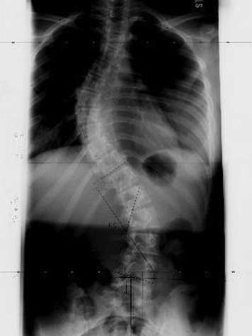
An X-ray is a type of radiation that passes through the body. It gives the surgeon information on the size, shape, and location of the bones in the spinal column. It is also called radiography.
By far, X-rays are the most common diagnostic tool in scoliosis. It tells the surgeon many things, most importantly, the size of your teenager’s curve.

Your teen, wearing a hospital gown, will be asked to stand next to the X-ray film. They will be asked to hold still for two or three seconds, so the picture doesn’t blur. The X-ray machine will be turned on for a fraction of a second. Beams of X-rays will pass through your teenager’s spine to make a picture on the film. The X-ray film, like regular camera film, is developed in about 10 minutes. In some hospitals, X-rays are completely digitized and your teen’s surgeon will view them directly on a computer.
Your teen won’t feel a thing. There is very little radiation released through the X-ray. To make it even more safe, the radiation technologist will provide shielding as needed. The type of shielding required depends on the type of X-ray being done. For example, because your teen is having spine X-rays, they will be given breast shields.
There are several types of X-rays that may be done on your teenager:
- Three-foot spinal posterior-anterior X-ray: This is an X-ray of your teen’s spine that is shot back to front.
- Three-foot lateral spinal X-ray: This is an X-ray of your teen’s spine that is shot from the side.
- Spinal bending films: These are X-rays of your teen’s spine to test its flexibility.
- Bone age X-ray: This is an X-ray of your teen’s left hand to assess whether they are still growing.
- Pelvis X-ray: This is an X-ray of your teen’s pelvis to assess whether their bones are still growing. X-rays of the elbow may also be done to assess bone maturity.