AboutKidsHealth


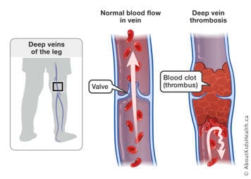
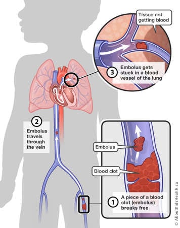
Deep vein thrombosisDDeep vein thrombosisDeep vein thrombosisEnglishHaematologyChild (0-12 years);Teen (13-18 years)NACardiovascular systemConditions and diseasesAdult (19+) CaregiversNA2017-09-25T04:00:00Z9.9000000000000056.30000000000002038.00000000000Health (A-Z) - ConditionsHealth A-Z<p>Learn what blood clots are, how they form, how they are treated, and complications that can occur because of them.</p><h2>​​What is deep vein thrombosis (DVT)?</h2><p>Deep vein thrombosis (DVT) is caused by a blood clot (thrombus) that occurs in the deep venous system. Deep veins are located within the muscles and are very important as they transport the blood back to the heart with the assistance of both vein valves and muscle contractions. Valves are found throughout the veins, especially in the legs, and help push the blood from the periphery of the body through the veins toward the heart. Hence, valves prevent backward blood flow, away from the heart. DVT can occur in the deep veins of the legs or arms. The blood clot fills the interior of the vein, obstructing the blood flow and causing several complications.</p> <figure class="asset-c-80"> <span class="asset-image-title">Deep vein thrombosis (DVT)</span> <img src="https://assets.aboutkidshealth.ca/AKHAssets/deep_vein_thrombosis_DVT_EN.jpg" alt="Side-by-side of normal blood flow in vein and deep vein thrombosis" /> <figcaption>Normally, blood flows easily through the deep veins of the body with the help of both vein valves and muscle contractions. Although less common than in adults, a child might also get a blood clot in a deep vein. The blood clot (thrombus) can fill the inside of the vein obstructing blood flow. </figcaption> </figure><h2>Key points</h2><ul><li>Deep vein thrombosis (DVT) is caused by a blood clot that occurs in the deep venous system.</li><li>DVTs are rare in healthy children, but may occur more commonly in children that are hospitalized. Children at higher risk include those with central lines, have a family history of increased clotting, or have certain anatomic variants that affect their veins.</li><li>Signs and symptoms of DVT include swelling, pain, and changes in skin colour of the affected limb.</li><li>Diagnosis of DVT is usually confirmed with an imaging test such as an ultrasound.</li><li>Treatment for DVT includes waiting and watching to see what happens, medication, thrombolysis, or surgery.</li><li>Rarely, DVT can cause a pulmonary embolism or a stroke. The most common chronic complication of DVT is known as post-thrombotic syndrome.</li></ul><h2>Signs and symptoms of DVT</h2> <p>DVTs located in the arms or the legs can be accompanied by limb swelling, pain, and changes in the color of the skin (red or bluish color).</p><h2>Causes of DVT</h2><p>DVTs are less common in children than in adults. Until recently, there was very little information describing the risk factors for DVT in children. However, some of the following risk factors found in adults are thought to also affect children who have DVT. Recent work by international research groups is helping to clarify risk factors for the development of DVT in children. Children admitted to a pediatric hospital are at the highest risk for thrombosis, which is largely due to the use of catheters or to their underlying health problem. </p><h3>Blood vessel damage</h3><p>DVT can be seen in children that have a central venous line or catheter that is used to give medications inside the deep venous system. In most cases, micro damage to the vein caused by the line causes platelets and clotting factors to start a clot. Blood also flows more slowly around the line. As a result, blood cells stick to the clot causing it to grow. </p><h3>Anatomic variants and exercise-induced DVT</h3><p>Changes in the anatomy or arrangement of blood vessels or the muscles close to blood vessels can lead to DVT in teens and young adults. These changes may cause tight corners within the blood vessels, which slow down the blood flow. Some anatomic changes can cause trauma to blood vessels during repetitive and intense exercise, resulting in blood clots. </p><h3>Medical and genetic conditions</h3><p>Certain infectious conditions (such as <a href="/article?contentid=2311&language=English">osteomyelitis</a>) and inflammatory conditions (such as antiphosphospholipid syndrome) can also trigger the clotting system. These conditions provide a false signal similar to the one that happens when an injury occurs, therefore increasing the risk of clot in the deep veins. </p><p>Similarly, conditions such as vasculitis (inflammation of the blood vessels) and some medications, such as chemotherapy, can lead to DVT.</p><p>In addition to medical conditions or medications, blood clots may appear in children whose blood clotting system produces clots more easily than those of other children. For example, some children may inherit genes from one or both of their parents that can increase their risk for developing a blood clot. In some cases, there can be a significant family history of clots, which present themselves in the form of DVT, heart attacks (myocardial infarction), strokes, or clots in the lungs (pulmonary embolism), and multiple miscarriages from clots in the placenta.</p><h3>Age</h3><p>As a person ages, the walls of their veins become less elastic and more susceptible to venous problems. Similarly, the circulating clotting factors rise with age, making clotting more common in older adults than in neonates and children.</p><h3>Immobility</h3><p>If a person is immobile (confined to a bed, unable to walk or spending large parts of the day in a bed or chair), the skeletal-muscle pumps, which help blood in the deep veins to return to the heart, are at rest. This leads to a slow blood flow inside of the deep veins of both legs, increasing the risk of blood clots. </p><p>In adults, there is a small risk of thrombosis when being relatively immobile when travelling by plane, train, car, bus or boat. The risk for DVT while travelling is higher for people affected by one or more of the other risk factors listed.</p><h3>Obesity</h3><p>Obesity is associated with conditions that may increase the risk of DVT. </p><h3>Dehydration</h3><p>When the body is dehydrated, the blood has a tendency to thicken, which increases the risk for developing DVT. </p><h3>Hormone therapy</h3><p>Birth control pills, patches or rings that contain estrogen increase the risk of DVT, particularly during the first year of usage.</p> <h2>Diagnosis of DVT</h2><p>Blood clots are suspected when the obstruction of a vein results in problems. For example, a leg with a blood clot in the deep veins becomes swollen, red, and painful. To confirm this suspicion, blood clots are usually diagnosed with an <a href="/article?contentid=1290&language=English">ultrasound</a>. An ultrasound is a medical test that uses sound waves to obtain images of structures inside the body, such as blood flow through a vein. In some cases, other imaging tests may be required. For example, to diagnose a clot in the lungs, a tomography (a special type of X-ray of the lungs) or a ventilation-perfusion scan (which measures the air and blood supply to the lungs) might be required. </p><p>In addition, children usually undergo blood tests when a clot is suspected to make sure it would be safe to start treatment, if required.</p><p>The thrombosis team is responsible for diagnosis, initial management, and follow up. The team will also coordinate your child’s future follow up in the thrombosis clinic, where the blood clot will continue to be monitored over time.<br></p><div class="asset-video"><iframe src="https://www.youtube.com/embed/l9VWSEqL6Zs"></iframe> </div><h2>Treatment of DVT</h2><p>There are four different treatments that can be done when a child is diagnosed with DVT.</p><h3>Wait and watch</h3><p>In some cases, your child’s doctor may decide not to treat the clot right away. This could happen if the clot is old, or if your child’s doctor determines that the risk of treatment outweighs the benefit. In those situations, it is reasonable not to give medications to help with the clot and to monitor your child closely. Your child’s doctor may take new imaging scans to evaluate if the clot is growing in the absence of treatment.</p><h3>Anticoagulant drugs</h3><p>Anticoagulants (blood thinners) can be prescribed to treat DVT. This treatment helps stabilize the clot, preventing growth and new clot formation so the body can use its own natural mechanisms to break down the clot. </p><p>These drugs include the anticoagulants from the family named heparinoids (standard heparin, <a href="/article?contentid=253&language=English">tinzaparin</a>, dalteparin, reviparin, nadroparin and <a href="/article?contentid=129&language=English">enoxaparin</a>), ultralow heparin (fondaparinux), or the family named oral <a href="/article?contentid=1937&language=English">vitamin K</a> antagonists (OVKA; <a href="/article?contentid=265&language=English">warfarin​</a>, acenocumarol, phenprocoumon). Heparinoids are administered through the veins (standard heparin) or as injections through the skin (subcutaneous injections; tinzaparin, dalteparin, reviparin, nadroparin, enoxaparin). Fondaparinux is also administered subcutaneously, whereas OVKA are administered by mouth. In all cases, anticoagulation medications in children due to a DVT are usually given for three months. Exceptions may occur for very young children (newborns and infants; six week-duration may be considered), or for patients that have a long-lasting thrombosis risk factor (duration longer than three months).<br></p><h3>Thrombolysis<br></h3><p>Thrombolysis is the process by which the clot is broken down either mechanically, or with the aid of a “clot-busting” medication. This option is considered only when there is an extensive blood clot, or when there is loss of blood supply to an organ or limb because of the clot’s presence, causing a risk of organ or limb loss. </p><p>Thrombolysis can be given in the hospital as an infusion, where the patient will be closely monitored. Thrombolysis may also be performed by an interventional radiologist using image guidance. The interventional radiologist uses a catheter to break up the blood clot, and monitors the procedure with X-ray imaging.</p><h3>Surgery</h3><p>In rare cases, surgery will be performed to remove the blood clot. Surgery as a treatment is rare and usually only used in emergency situations.<br></p><h2>Complications of blood clots and DVT</h2><p>Fresh clots like to grow within the deep venous system. Occasionally, a smaller portion of the clot breaks off and is transported into different locations of the body, depending on where it originated. Sometimes, the fragmented blood clot travels all the way to the lungs. When this happens, it is called a pulmonary embolism. </p> <figure class="asset-c-80"> <span class="asset-image-title">Pulmonary embolism</span><img src="https://assets.aboutkidshealth.ca/AKHAssets/pulmonary_embolism_EN_XL.jpg" alt="Piece of a blood clot breaking free in the leg, traveling through the vein and getting stuck in a blood vessel in the lung" /><figcaption>1) A small piece of a fresh blood clot in a vein can break free (embolus). 2) The embolus travels through the veins of the body to the heart and into the lung. 3) The embolus gets stuck in a blood vessel in the lung. This blocks blood flow to a part of the lung.</figcaption> </figure> <p>Very rarely, when a small hole in the heart is present, the blood flow of the right side of the heart may travel to the left side. This allows the blood clot to enter the left side of the heart, which provides blood to the brain. This increases the risk of a paradoxical embolism or stroke.</p><p>Other potential complications of clots are recurrence (when the clot comes back or there is a new clot somewhere else) and <a href="/Article?contentid=2884&language=English">post-thrombotic syndrome</a>. </p><p>It is highly unlikely that part of a blood clot left in the vessel after some time will travel to a new location in the body. After about six weeks, the clot starts to become “old” and calcifies, becoming part of the vessel. At this point the clot is considered to be stable and unlikely to cause further damage.</p><h2>At SickKids</h2><p>​​The thrombosis team at SickKids includes a nurse practitioner (NP), staff physicians, thrombosis fellows, and research staff.</p><p>If you are a SickKids patient, you will attend follow-up appointments in the thrombosis out-patient clinic, open Mondays, Wednesdays, and Fridays.</p><p>For SickKids patients, please see below for contact information in non-urgent and urgent situations:</p><ul><li>For non-urgent clinical matters, contact the Nurse Practitioner at: 416-813-8514</li> <li>For appointment clarification or rescheduling, contact the Thrombosis Clinic Coordinator at: 416-813-5453 extension 2</li><li>For after-hours clinical emergencies, contact the Thrombosis Fellow on-call at: 416-813-7500</li></ul><p>For more information on thrombosis, post-thrombotic syndrome and the management of these conditions, please visit the <a href="/thrombosis">Thrombosis Learning Hub</a>.<br></p>
Thrombose veineuse profondeTThrombose veineuse profondeDeep vein thrombosisFrenchHaematologyChild (0-12 years);Teen (13-18 years)NACardiovascular systemConditions and diseasesAdult (19+) CaregiversNA2017-09-25T04:00:00ZHealth (A-Z) - ConditionsHealth A-Z<p>Découvrez ce que sont les caillots sanguins, de quelle façon ils se forment, de quelle façon ils sont traités et les complications qu’ils peuvent causer.</p><h2>​​Qu’est-ce qu’une thrombose veineuse profonde (TVP)?</h2><p>Une thrombose veineuse profonde (TVP) est causée par un caillot sanguin (thrombus) qui se forme dans le système veineux profond. Les veines profondes sont situées à l’intérieur des muscles et sont très importantes, car elles renvoient le sang vers le cœur grâce aux valvules veineuses et des contractions musculaires. Les valvules sont distribuées tout au long des veines, particulièrement dans les jambes, et elles aident à pousser le sang de la périphérie de l’organisme vers le cœur. De ce fait, les valvules préviennent le reflux sanguin, en direction opposée au cœur. La thrombose veineuse pr​ofonde peut se produire dans les veines profondes des jambes ou des bras. Le caillot sanguin remplit l’intérieur de la veine, obstruant la circulation sanguine et causant plusieurs complications.</p> <figure class="asset-c-80"> <span class="asset-image-title">Thrombose veineuse profonde (TVP)</span> <img src="https://assets.aboutkidshealth.ca/AKHAssets/deep_vein_thrombosis_DVT_FR.jpg" alt="Illustration de la circulation sanguine normale dans la veine et de la thrombose veineuse profonde" /> <figcaption>En temps normal, le sang circule facilement dans les veines profondes du corps, grâce aux valvules veineuses et aux contractions musculaires. Même si cela est moins fréquent que chez l’adulte, un caillot sanguin dans une veine profonde peut se former chez l’enfant. Le caillot sanguin (thrombus) peut remplir l’intérieur de la veine et obstruer la circulation sanguine.</figcaption> </figure><h2>À retenir</h2><ul><li>Une thrombose veineuse profonde (TVP) est causée par un caillot sanguin qui se forme dans le système veineux profond.</li><li>Les thromboses veineuses profondes sont rares chez les enfants en bonne santé, mais peuvent survenir plus fréquemment chez les enfants qui sont hospitalisés. Les enfants présentant un risque élevé comprennent ceux avec des voies veineuses centrales, ceux qui présentent des antécédents familiaux d’augmentation de la coagulation sanguine ou qui ont certaines particularités anatomiques qui affectent leurs veines.</li><li>Les symptômes de la thrombose veineuse profonde (TVP) comprennent de l’enflure, de la douleur et une décoloration de la peau du membre atteint.</li><li>Le diagnostic de la thrombose veineuse profonde est confirmé par un examen d’imagerie comme une échographie.</li><li>Le traitement de la thrombose veineuse profonde comprend d’observer et d’attendre afin de suivre l’évolution, des médicaments, une thrombolyse ou une intervention chirurgicale.</li><li>Dans de rares cas, la thrombose veineuse profonde peut causer une embolie pulmonaire ou accident vasculaire cérébral. La complication chronique la plus fréquente de la thrombose veineuse profonde est la maladie post-phlébitique.</li></ul><h2>Symptômes de la thrombose veineuse profonde (TVP)</h2> <p>La thrombose veineuse profonde située dans les bras ou les jambes peut être accompagnée par une enflure du membre, de la douleur et une décoloration de la peau (couleur rouge ou bleutée).</p><h2>Causes de la thrombose veineuse profonde (TVP)</h2><p>Les thromboses veineuses profondes (TVP) sont moins fréquentes chez les enfants que chez les adultes. Jusqu’à récemment, il existait très peu de documentation décrivant les facteurs de risques de la thrombose veineuse profonde chez les enfants. Cependant, on croit que certains des facteurs de risques suivants observés chez les adultes touchent aussi les enfants atteints de thrombose veineuse profonde. Des travaux récents, réalisés par des groupes de recherche internationaux, contribuent à clarifier les facteurs de risques de l’évolution de la thrombose veineuse profonde chez les enfants. Les enfants admis à un hôpital pédiatrique sont les plus à risques d’être atteint d’une thrombose, laquelle est surtout causée par l’emploi de cathéters ou d’un problème de santé sous-jacent.</p><h3>Lésions aux vaisseaux sanguins</h3><p>Une thrombose veineuse profonde peut être observée chez les enfants qui ont une voie veineuse centrale ou un cathéter employé dans le but d’administrer des médicaments au sein du système veineux profond. Dans la plupart des cas, des lésions microscopiques causées aux veines par la voie veineuse entraînent les plaquettes et les facteurs de coagulation à former un caillot. Le sang circule aussi plus lentement autour des voies. Il en résulte que les cellules sanguines s’agglomèrent au caillot et contribuent à sa croissance.</p><h3>Variantes anatomiques de la thrombose veineuse profonde, causées ou non par l’exercice</h3><p>Des changements dans l’anatomie ou la distribution des vaisseaux sanguins ou encore des muscles situés près des vaisseaux sanguins peuvent induire des thromboses veineuses profondes chez les adolescents et les jeunes adultes. Ces changements peuvent engendrer des coudes serrés à l’intérieur des vaisseaux sanguins, contribuant à ralentir la circulation sanguine. Certains changements anatomiques peuvent causer des traumatismes aux vaisseaux sanguins lors de la pratique d’exercices intenses et répétitifs, créant des caillots.</p><h3>Troubles médicaux et génétiques<br></h3><p>Certains troubles infectieux (tel que l’<a href="/Article?contentid=2311&language=French">ostéomyélite</a>) et inflammatoires (comme le syndrome des antiphospholipides) peuvent aussi déclencher le système de coagulation. Ces troubles déclenchent un faux signal, similaire à celui se produisant lors d’une blessure, augmentant ainsi le risque de caillot dans les veines profondes.</p><p>De manière similaire, des troubles comme la vascularite (une inflammation des vaisseaux sanguins) et certains médicaments, comme la chimiothérapie, peuvent mener à une thrombose veineuse profonde.</p><p>En plus des troubles médicaux ou des médicaments, des caillots sanguins peuvent survenir chez les enfants dont le système de coagulation sanguine produit des caillots plus facilement que celui des autres enfants. Par exemple, certains enfants peuvent hériter de gènes de l’un leurs parents, ou des deux, qui peuvent augmenter le risque de former un caillot sanguin. Dans certains cas, il peut y avoir des antécédents familiaux importants de caillots sanguins, lesquels se présentent sous la forme de thrombose veineuse profonde, de crises cardiaques (infarctus du myocarde), d’accidents vasculaires cérébraux ou de caillots sanguins dans les poumons (embolie pulmonaire) ainsi que de multiples fausses-couches causées par des caillots dans le placenta.</p><h3>Âge</h3><p>Plus une personne vieillit, plus les parois de ses veines perdent en élasticité et deviennent plus susceptibles aux problèmes veineux. De manière similaire, la présence de facteurs de coagulation en circulation augmente avec l’âge, rendant les caillots sanguins plus fréquents chez les adultes plus âgés que chez les nouveau-nés et les enfants.</p><h3>Immobilité</h3><p>Lorsqu’une personne est immobilisée (confinée à un lit, incapable de marcher ou passant une bonne partie de ses journées au lit ou dans un fauteuil), les pompes musculaires squelettiques, qui aident le sang contenu dans les veines profondes à retourner au cœur, sont au repos. Ceci entraîne un ralentissement de la circulation sanguine au sein des veines profondes des jambes, augmentant le risque de caillots sanguins.</p><p>Chez les adultes, il existe un faible risque de thrombose lorsqu’une personne reste relativement immobile au cours d’un voyage en avion, en train, en voiture, en autobus ou en bateau. Le risque de thrombose veineuse profonde lors de déplacement est plus élevé chez les personnes touchées par un ou plusieurs autres facteurs de risques parmi ceux listés ici.</p><h3>Obésité</h3><p>L’obésité est associée à des troubles qui peuvent augmenter le risque de thrombose veineuse profonde.</p><h3>Déshydratation</h3><p>Lorsque le corps est déshydraté, le sang a tendance à devenir plus épais, ce qui augmente le risque de thrombose veineuse profonde.</p><h3>Thérapie hormonale</h3><p>Les pilules contraceptives, les timbres ou les anneaux qui contiennent de l’œstrogène augmentent le risque de thrombose veineuse profonde, particulièrement pendant la première année d’utilisation. Il existe aussi un risque accru de caillots sanguins au cours de la grossesse. Par conséquent, consulter un professionnel en soins de santé concernant les méthodes de contraception les mieux adaptées est très important, surtout pour les adolescentes présentant des problèmes de santé antérieurs ou pour celles qui présentent des antécédents familiaux de caillots sanguins.</p><h2>Diagnostic de la thrombose veineuse profonde</h2><p>On suspecte la présence de caillots sanguins lorsque l’obstruction d’une veine entraîne des problèmes. Par exemple, une jambe présentant un caillot sanguin au sein d’une veine profonde deviendra enflée, rouge et douloureuse. Afin de confirmer ce soupçon, les caillots sanguins sont habituellement diagnostiqués à l’aide d’une <a href="/article?contentid=1290&language=French">échographie</a>. Une échographie est un examen médical qui emploie les ondes sonores afin d’obtenir des images des structures situées à l’intérieur du corps, comme la circulation sanguine au travers d’une veine. Dans certains cas, d’autres examens d’imagerie pourraient être nécessaires. Par exemple, une tomographie (un type particulier de radiographie des poumons) ou une scintigraphie pulmonaire de ventilation-perfusion (qui mesure l’approvisionnement des poumons en air et en sang) pourrait être nécessaire afin de diagnostiquer un caillot sanguin logé dans les poumons.</p><p>De plus, les enfants sont généralement soumis à des analyses sanguines lorsqu’on suspecte la présence d’un caillot sanguin afin de s’assurer qu’on puisse débuter le traitement de manière sécuritaire, si nécessaire.</p><p>L’équipe de thrombose est responsable du diagnostic, de la gestion initiale et du suivi. L’équipe coordonnera aussi les prochains rendez-vous de suivi de l’enfant à la clinique de thrombose, où la surveillance du caillot sanguin se poursuivra dans le temps.</p><h2>Traitement de la thrombose veineuse profonde</h2><p>Lorsqu’un enfant obtient un diagnostic de thrombose veineuse profonde, quatre traitements différents peuvent être employés.</p><h3>Observer et attendre</h3><p>Dans certains cas, le médecin de votre enfant pourrait décider de ne pas traiter immédiatement le caillot sanguin. Cela pourrait arriver si le caillot est ancien ou si le médecin de votre enfant détermine que le risque est plus grand que les avantages associés au traitement. Dans ces situations, il est raisonnable de ne pas donner de médicaments contre le caillot et de surveiller votre enfant de près. Le médecin de votre enfant pourrait procéder à de nouveaux examens d’imagerie afin d’évaluer si le caillot sanguin grossit en absence de traitement.</p><h3>Médicaments anticoagulants</h3><p>Des anticoagulants (ou fluidifiants sanguins) peuvent être prescrits afin de traiter la thrombose veineuse profonde. Ce traitement aide à stabiliser le caillot, à prévenir sa croissance et la formation de nouveaux caillots de telle sorte que le corps peut le détruire en employant ses mécanismes naturels.</p><p>Ces médicaments comprennent des anticoagulants de la famille des héparinoïdes (héparine standard, <a href="/article?contentid=253&language=French">tinzaparin</a>, daltéparine, réviparine, nardroparine et l’<a href="/article?contentid=129&language=French">énoxaparine</a>), de l’héparine de très faible poids moléculaire (Fondaparinux) ou de la famille des antagonistes de la <a href="/article?contentid=1937&language=French">vitamine K</a> administrés sous forme orale (AVK, <a href="/article?contentid=265&language=French">warfarine</a>, acénocoumarol, phénprocoumone). Les héparinoïdes sont administrés par voie veineuse (standard héparine) ou injectés à travers la peau (injections sous-cutanées); tinzaparine, daltéparine, réviparine, nardroparine et énoxaparine). Le Fondaparinux est aussi administré de manière sous-cutanée alors que l'AVK est administré par la bouche. Dans tous les cas, les médicaments anticoagulants destinés aux enfants atteints de thrombose veineuse profonde sont habituellement prescrits pour trois mois. Il peut exister des exceptions chez les très jeunes enfants (nouveau-nés, nourrissons; une période de six semaines peut être prise en considération) ou pour les patients présentant des facteurs de risques de thrombose à long terme (d’une durée supérieure à trois mois).</p><h3>Thrombolyse</h3><p>La thrombolyse est le processus par lequel un caillot sanguin est désagrégé, soit de manière mécanique ou à l’aide d’un médicament « briseur de caillot ». Cette option est prise en considération uniquement lorsque le caillot sanguin est étendu ou lorsque sa présence compromet l’approvisionnement en sang d’un organe ou d’un membre au point où il y a un risque de perdre l’organe ou le membre.</p><p>La thrombolyse peut être administrée sous forme d’infusion à l’hôpital, là où le patient sera étroitement surveillé. La thrombolyse peut aussi être réalisée par un radiologue d’intervention guidé par images. Le radiologue d’intervention utilise un cathéter afin de briser le caillot sanguin et il suit la procédure à l’aide d’imagerie rayons X.</p><h3>Intervention chirurgicale</h3><p>Dans de rares cas, une intervention chir</p><h2>Complications des caillots sanguins et de la thrombose veineuse profonde</h2><p>Les nouveaux caillots ont tendance à croître au sein du système veineux profond. À l’occasion, une petite partie du caillot sanguin se déloge et elle est transportée dans une autre partie du corps, selon l’endroit d’où elle provient. Parfois, un fragment de caillot sanguin se déplace jusqu’aux poumons. Lorsque cela se produit, on parle d’une embolie pulmonaire.</p> <figure class="asset-c-80"> <span class="asset-image-title">Embolie pulmonaire</span> <img src="https://assets.aboutkidshealth.ca/AKHAssets/pulmonary_embolism_FR_XL.jpg" alt="Partie d’un caillot sanguin se libérant, se déplaçant le long de la veine et se coinçant dans un vaisseau sanguin du poumon" /> <figcaption>1) Une petite partie d’un caillot sanguin fraîchement formé dans une veine peut se libérer (un embole). 2) L’embole se déplace dans les veines jusqu’au cœur puis dans le poumon. 3) L’embole reste coincé dans un vaisseau sanguin du poumon, bloquant le flux sanguin vers une partie du poumon.</figcaption> </figure> <p>Très rarement, lorsqu’un petit trou est présent dans le cœur, le sang qui circule dans le côté droit du cœur peut se déplacer dans le côté gauche. Le caillot sanguin peut alors pénétrer dans le côté gauche du cœur, lequel approvisionne le cerveau en sang. Ceci augmente le risque d’embolie paradoxale ou d’accident vasculaire cérébral.</p><p>D’autres complications potentielles liées à la présence de caillot sanguin sont la récidive (lorsqu’un caillot se reforme ou qu’un nouveau caillot apparaît à un autre endroit) et la <a href="/article?contentid=2884&language=French">maladie post-phlébitique</a>.</p><p>Il est très peu probable qu’une partie du caillot sanguin laissée dans le vaisseau pour un certain temps se déplacera à un nouvel endroit dans le corps. Après environ six semaines, le caillot sanguin commence à « vieillir » et à se calcifier, devenant partie intégrante du vaisseau sanguin. Rendu à ce point, le caillot sanguin est considéré comme stable et peu susceptible de causer d’autres dommages.</p><h2>À l’hôpital SickKids</h2><p>L’équipe de thrombose de l’hôpital SickKids comprend un infirmier praticien, des médecins traitants, des étudiants se spécialisant en thrombose et du personnel de recherche. Si vous êtes un patient à SickKids, vous assisterez à des rendez-vous de suivi à notre clinique externe de thrombose, ouverte les lundis, mercredis et vendredis.</p><p>Pour les patients de SickKids, veuillez consulter les renseignements suivants dans les situations non urgentes et urgentes :</p><ul><li>pour les questions cliniques non urgentes, communiquez avec l’infirmier praticien au : 416-813-8514</li><li>pour des renseignements sur votre rendez-vous ou pour le modifier, veuillez communiquer avec le coordonnateur de la clinique de thrombose au : 416 813-5453, poste 2</li><li>pour les urgences cliniques survenant en dehors des heures d’ouverture, communiquez avec le médecin de garde au : 416-813-7500</li></ul>


Deep vein thrombosis2534.00000000000Deep vein thrombosisDeep vein thrombosisDEnglishHaematologyChild (0-12 years);Teen (13-18 years)NACardiovascular systemConditions and diseasesAdult (19+) CaregiversNA2017-09-25T04:00:00Z9.9000000000000056.30000000000002038.00000000000Health (A-Z) - ConditionsHealth A-Z<p>Learn what blood clots are, how they form, how they are treated, and complications that can occur because of them.</p><h2>​​What is deep vein thrombosis (DVT)?</h2><p>Deep vein thrombosis (DVT) is caused by a blood clot (thrombus) that occurs in the deep venous system. Deep veins are located within the muscles and are very important as they transport the blood back to the heart with the assistance of both vein valves and muscle contractions. Valves are found throughout the veins, especially in the legs, and help push the blood from the periphery of the body through the veins toward the heart. Hence, valves prevent backward blood flow, away from the heart. DVT can occur in the deep veins of the legs or arms. The blood clot fills the interior of the vein, obstructing the blood flow and causing several complications.</p> <figure class="asset-c-80"> <span class="asset-image-title">Deep vein thrombosis (DVT)</span> <img src="https://assets.aboutkidshealth.ca/AKHAssets/deep_vein_thrombosis_DVT_EN.jpg" alt="Side-by-side of normal blood flow in vein and deep vein thrombosis" /> <figcaption>Normally, blood flows easily through the deep veins of the body with the help of both vein valves and muscle contractions. Although less common than in adults, a child might also get a blood clot in a deep vein. The blood clot (thrombus) can fill the inside of the vein obstructing blood flow. </figcaption> </figure><h3>What are blood clots?</h3> <p>Clotting factors are proteins in the blood that help to stop cuts and wounds from bleeding. Platelets, one of main elements in blood, work together with the clotting factors to stop bleeding by creating blood clots. Blood clots are clumps of blood, including clotting factors and platelets, which harden over time to stop wounds from bleeding.</p><h2>Key points</h2><ul><li>Deep vein thrombosis (DVT) is caused by a blood clot that occurs in the deep venous system.</li><li>DVTs are rare in healthy children, but may occur more commonly in children that are hospitalized. Children at higher risk include those with central lines, have a family history of increased clotting, or have certain anatomic variants that affect their veins.</li><li>Signs and symptoms of DVT include swelling, pain, and changes in skin colour of the affected limb.</li><li>Diagnosis of DVT is usually confirmed with an imaging test such as an ultrasound.</li><li>Treatment for DVT includes waiting and watching to see what happens, medication, thrombolysis, or surgery.</li><li>Rarely, DVT can cause a pulmonary embolism or a stroke. The most common chronic complication of DVT is known as post-thrombotic syndrome.</li></ul><h2>Signs and symptoms of DVT</h2> <p>DVTs located in the arms or the legs can be accompanied by limb swelling, pain, and changes in the color of the skin (red or bluish color).</p><h2>Causes of DVT</h2><p>DVTs are less common in children than in adults. Until recently, there was very little information describing the risk factors for DVT in children. However, some of the following risk factors found in adults are thought to also affect children who have DVT. Recent work by international research groups is helping to clarify risk factors for the development of DVT in children. Children admitted to a pediatric hospital are at the highest risk for thrombosis, which is largely due to the use of catheters or to their underlying health problem. </p><h3>Blood vessel damage</h3><p>DVT can be seen in children that have a central venous line or catheter that is used to give medications inside the deep venous system. In most cases, micro damage to the vein caused by the line causes platelets and clotting factors to start a clot. Blood also flows more slowly around the line. As a result, blood cells stick to the clot causing it to grow. </p><h3>Anatomic variants and exercise-induced DVT</h3><p>Changes in the anatomy or arrangement of blood vessels or the muscles close to blood vessels can lead to DVT in teens and young adults. These changes may cause tight corners within the blood vessels, which slow down the blood flow. Some anatomic changes can cause trauma to blood vessels during repetitive and intense exercise, resulting in blood clots. </p><h3>Medical and genetic conditions</h3><p>Certain infectious conditions (such as <a href="/article?contentid=2311&language=English">osteomyelitis</a>) and inflammatory conditions (such as antiphosphospholipid syndrome) can also trigger the clotting system. These conditions provide a false signal similar to the one that happens when an injury occurs, therefore increasing the risk of clot in the deep veins. </p><p>Similarly, conditions such as vasculitis (inflammation of the blood vessels) and some medications, such as chemotherapy, can lead to DVT.</p><p>In addition to medical conditions or medications, blood clots may appear in children whose blood clotting system produces clots more easily than those of other children. For example, some children may inherit genes from one or both of their parents that can increase their risk for developing a blood clot. In some cases, there can be a significant family history of clots, which present themselves in the form of DVT, heart attacks (myocardial infarction), strokes, or clots in the lungs (pulmonary embolism), and multiple miscarriages from clots in the placenta.</p><h3>Age</h3><p>As a person ages, the walls of their veins become less elastic and more susceptible to venous problems. Similarly, the circulating clotting factors rise with age, making clotting more common in older adults than in neonates and children.</p><h3>Immobility</h3><p>If a person is immobile (confined to a bed, unable to walk or spending large parts of the day in a bed or chair), the skeletal-muscle pumps, which help blood in the deep veins to return to the heart, are at rest. This leads to a slow blood flow inside of the deep veins of both legs, increasing the risk of blood clots. </p><p>In adults, there is a small risk of thrombosis when being relatively immobile when travelling by plane, train, car, bus or boat. The risk for DVT while travelling is higher for people affected by one or more of the other risk factors listed.</p><h3>Obesity</h3><p>Obesity is associated with conditions that may increase the risk of DVT. </p><h3>Dehydration</h3><p>When the body is dehydrated, the blood has a tendency to thicken, which increases the risk for developing DVT. </p><h3>Hormone therapy</h3><p>Birth control pills, patches or rings that contain estrogen increase the risk of DVT, particularly during the first year of usage.</p> <h2>Diagnosis of DVT</h2><p>Blood clots are suspected when the obstruction of a vein results in problems. For example, a leg with a blood clot in the deep veins becomes swollen, red, and painful. To confirm this suspicion, blood clots are usually diagnosed with an <a href="/article?contentid=1290&language=English">ultrasound</a>. An ultrasound is a medical test that uses sound waves to obtain images of structures inside the body, such as blood flow through a vein. In some cases, other imaging tests may be required. For example, to diagnose a clot in the lungs, a tomography (a special type of X-ray of the lungs) or a ventilation-perfusion scan (which measures the air and blood supply to the lungs) might be required. </p><p>In addition, children usually undergo blood tests when a clot is suspected to make sure it would be safe to start treatment, if required.</p><p>The thrombosis team is responsible for diagnosis, initial management, and follow up. The team will also coordinate your child’s future follow up in the thrombosis clinic, where the blood clot will continue to be monitored over time.<br></p><div class="asset-video"><iframe src="https://www.youtube.com/embed/l9VWSEqL6Zs"></iframe> </div><h2>Treatment of DVT</h2><p>There are four different treatments that can be done when a child is diagnosed with DVT.</p><h3>Wait and watch</h3><p>In some cases, your child’s doctor may decide not to treat the clot right away. This could happen if the clot is old, or if your child’s doctor determines that the risk of treatment outweighs the benefit. In those situations, it is reasonable not to give medications to help with the clot and to monitor your child closely. Your child’s doctor may take new imaging scans to evaluate if the clot is growing in the absence of treatment.</p><h3>Anticoagulant drugs</h3><p>Anticoagulants (blood thinners) can be prescribed to treat DVT. This treatment helps stabilize the clot, preventing growth and new clot formation so the body can use its own natural mechanisms to break down the clot. </p><p>These drugs include the anticoagulants from the family named heparinoids (standard heparin, <a href="/article?contentid=253&language=English">tinzaparin</a>, dalteparin, reviparin, nadroparin and <a href="/article?contentid=129&language=English">enoxaparin</a>), ultralow heparin (fondaparinux), or the family named oral <a href="/article?contentid=1937&language=English">vitamin K</a> antagonists (OVKA; <a href="/article?contentid=265&language=English">warfarin​</a>, acenocumarol, phenprocoumon). Heparinoids are administered through the veins (standard heparin) or as injections through the skin (subcutaneous injections; tinzaparin, dalteparin, reviparin, nadroparin, enoxaparin). Fondaparinux is also administered subcutaneously, whereas OVKA are administered by mouth. In all cases, anticoagulation medications in children due to a DVT are usually given for three months. Exceptions may occur for very young children (newborns and infants; six week-duration may be considered), or for patients that have a long-lasting thrombosis risk factor (duration longer than three months).<br></p><h3>Thrombolysis<br></h3><p>Thrombolysis is the process by which the clot is broken down either mechanically, or with the aid of a “clot-busting” medication. This option is considered only when there is an extensive blood clot, or when there is loss of blood supply to an organ or limb because of the clot’s presence, causing a risk of organ or limb loss. </p><p>Thrombolysis can be given in the hospital as an infusion, where the patient will be closely monitored. Thrombolysis may also be performed by an interventional radiologist using image guidance. The interventional radiologist uses a catheter to break up the blood clot, and monitors the procedure with X-ray imaging.</p><h3>Surgery</h3><p>In rare cases, surgery will be performed to remove the blood clot. Surgery as a treatment is rare and usually only used in emergency situations.<br></p><h2>Complications of blood clots and DVT</h2><p>Fresh clots like to grow within the deep venous system. Occasionally, a smaller portion of the clot breaks off and is transported into different locations of the body, depending on where it originated. Sometimes, the fragmented blood clot travels all the way to the lungs. When this happens, it is called a pulmonary embolism. </p> <figure class="asset-c-80"> <span class="asset-image-title">Pulmonary embolism</span><img src="https://assets.aboutkidshealth.ca/AKHAssets/pulmonary_embolism_EN_XL.jpg" alt="Piece of a blood clot breaking free in the leg, traveling through the vein and getting stuck in a blood vessel in the lung" /><figcaption>1) A small piece of a fresh blood clot in a vein can break free (embolus). 2) The embolus travels through the veins of the body to the heart and into the lung. 3) The embolus gets stuck in a blood vessel in the lung. This blocks blood flow to a part of the lung.</figcaption> </figure> <p>Very rarely, when a small hole in the heart is present, the blood flow of the right side of the heart may travel to the left side. This allows the blood clot to enter the left side of the heart, which provides blood to the brain. This increases the risk of a paradoxical embolism or stroke.</p><p>Other potential complications of clots are recurrence (when the clot comes back or there is a new clot somewhere else) and <a href="/Article?contentid=2884&language=English">post-thrombotic syndrome</a>. </p><p>It is highly unlikely that part of a blood clot left in the vessel after some time will travel to a new location in the body. After about six weeks, the clot starts to become “old” and calcifies, becoming part of the vessel. At this point the clot is considered to be stable and unlikely to cause further damage.</p><h2>Prevention of blood clots and DVT</h2> <p>The best way to prevent blood clots and DVT is to maintain a healthy lifestyle. </p><ul><li>Exercise: Going on daily walks or participating in other aerobic exercises such as swimming can improve blood circulation and prevent new clots from forming.</li><li>Stay hydrated: Drinking plenty of water reduces risks of blood clotting by maintaining a liquefied consistency in the blood. Avoid substances that dehydrate the body, such as caffeine and alcohol.</li><li>Avoid smoking: Smoking increases the likelihood of platelets sticking together and damages the lining of the blood vessels. If you or another family member smokes, and your child is at risk for blood clots, make sure you keep your home, vehicle and anywhere else your child will be, a smoke-free environment.</li></ul><h2>At SickKids</h2><p>​​The thrombosis team at SickKids includes a nurse practitioner (NP), staff physicians, thrombosis fellows, and research staff.</p><p>If you are a SickKids patient, you will attend follow-up appointments in the thrombosis out-patient clinic, open Mondays, Wednesdays, and Fridays.</p><p>For SickKids patients, please see below for contact information in non-urgent and urgent situations:</p><ul><li>For non-urgent clinical matters, contact the Nurse Practitioner at: 416-813-8514</li> <li>For appointment clarification or rescheduling, contact the Thrombosis Clinic Coordinator at: 416-813-5453 extension 2</li><li>For after-hours clinical emergencies, contact the Thrombosis Fellow on-call at: 416-813-7500</li></ul><p>For more information on thrombosis, post-thrombotic syndrome and the management of these conditions, please visit the <a href="/thrombosis">Thrombosis Learning Hub</a>.<br></p>https://assets.aboutkidshealth.ca/AKHAssets/deep_vein_thrombosis_DVT_EN.jpgDeep vein thrombosisFalse

Thank you to our sponsors

AboutKidsHealth is proud to partner with the following sponsors as they support our mission to improve the health and wellbeing of children in Canada and around the world by making accessible health care information available via the internet.

Our Sponsors