AboutKidsHealth


The normal heartTThe normal heartThe normal heartEnglishCardiologyChild (0-12 years);Teen (13-18 years);Adult (19+)HeartCardiovascular systemNAAdult (19+)NA2021-01-28T05:00:00Z6.4000000000000071.90000000000001262.00000000000Flat ContentHealth A-Z<p>The heart is a muscle that pumps blood through the body. Learn about each part of the heart including what each part looks like, its specific function and its location.<br></p><h2>What is the heart?</h2><p><a href="/body/interactive?module=heart">The heart</a> is a muscle just a bit bigger than the size of your fist. It is located between the lungs, almost in the middle of the chest. It is the body's core muscle and the very first organ to form in the body after conception.</p><h2> Key points </h2><ul><li> The heart is located between the lungs and pumps blood throughout the body's cardiovascular system.</li><li>The heart has four chambers. The upper two chambers are the right atrium and left atrium and the lower two chambers are the right ventricle and left ventricle. The right and left sides of the heart are divided by a wall called the septum.</li><li>You can feel your pulse each time the ventricle pumps by feeling your wrist or side of your neck.</li></ul>
Un aperçu du cœurUUn aperçu du cœurThe normal heartFrenchCardiologyChild (0-12 years);Teen (13-18 years);Adult (19+)HeartCardiovascular systemNAAdult (19+)NA2009-12-04T05:00:00Z7.0000000000000071.0000000000000741.000000000000Flat ContentHealth A-Z<p>Découvrez les parties du corps et l’aspect de chacune de ces parties, leur fonction précise et leur emplacement.<br></p><p>Le cœur est un muscle tout juste plus gros que la taille de votre poing. Il est situé entre les poumons, presque au centre de votre poitrine. C’est le muscle central du corps, et le tout premier organe à se former après la conception.</p><h2> À retenir </h2> <ul><li> Le cœur se trouve entre les poumons et pompe le sang dans tout l’appareil cardiovasculaire du corps.</li> <li> Les côtés droit et gauche du cœur sont divisés par une paroi appelée le septum. Les deux cavités supérieures sont l’atrium droit et l’atrium gauche, tandis que les deux cavités inférieures sont le ventricule droit et le ventricule gauche.</li> <li> Vous pouvez sentir votre pouls chaque fois que le ventricule se contracte en vous tâtant le poignet ou le côté du cou.</li></ul>


Heart conditions: CongenitalHeart conditions: CongenitalHeart conditions: CongenitalHEnglishCardiologyChild (0-12 years);Teen (13-18 years)HeartCardiovascular systemConditions and diseasesAdult (19+) CaregiversNA2018-03-15T04:00:00ZLanding PageCollection Hub<p>Congenital heart conditions are present at birth. They are defects that are caused when parts of the heart do not form properly. They occur very early in a pregnancy, often even before a woman knows she is pregnant.</p><p>Congenital heart conditions are present at birth. They are defects that are caused when parts of the heart do not form properly. They occur very early in a pregnancy, often even before a woman knows she is pregnant. These defects can include holes in parts of the heart, or missing or malformed parts of the heart.</p><div class="panel panel-primary"><div class="panel-heading clickable"> <span class="pull-right panel-heading-collapsable-icon"><i class="mdi mdi-chevron-down"></i></span> <h2 class="panel-title">The heart and the circulatory system</h2></div><div class="panel-body list-group" style="display:none;"><p>The heart is a vital organ that sits between the lungs in your chest. It pumps blood around the body through a network a blood vessels (arteries and veins). Learn about the heart’s anatomy, its role in the body and how it works.</p></div><ol class="list-group" style="display:none;"><li class="list-group-item"> <a class="overview-links" href="/Article?contentid=1577&language=English">The normal heart</a></li><li class="list-group-item"> <a class="overview-links" href="/Article?contentid=1579&language=English">The circulatory system before and after birth</a></li><li class="list-group-item"> <a class="overview-links" href="/Article?contentid=1578&language=English">Blood</a></li></ol></div><div class="panel panel-primary"><div class="panel-heading clickable"> <span class="pull-right panel-heading-collapsable-icon"><i class="mdi mdi-chevron-down"></i></span> <h2 class="panel-title">Aortic atresia</h2></div><div class="panel-body list-group" style="display:none;"><p>Learn about aortic atresia where there is no opening from the left ventricle of the heart into the aorta. Read about some of the tests and procedures used to diagnose and treat the condition. </p></div><ol class="list-group" style="display:none;"><li><div class="panel-heading clickable"> <span class="pull-right panel-heading-collapsable-icon"><i class="mdi mdi-chevron-down"></i></span> <h3>Overview</h3></div><ol class="list-group" style="display:none;"><li class="list-group-item"> <a class="overview-links" href="/Article?contentid=1605&language=English">Aortic atresia</a></li></ol></li><li><div class="panel-heading clickable"> <span class="pull-right panel-heading-collapsable-icon"><i class="mdi mdi-chevron-down"></i></span> <h3>Tests and diagnosis</h3></div><ol class="list-group" style="display:none;"><li class="list-group-item"> <a class="overview-links" href="/Article?contentid=1647&language=English">Imaging tests: X-ray, MRI and CT scan</a></li><li class="list-group-item"> <a class="overview-links" href="/Article?contentid=1276&language=English">Electrocardiogram (ECG) test</a></li><li class="list-group-item"> <a class="overview-links" href="/Article?contentid=1274&language=English">Echocardiogram</a></li><li class="list-group-item"> <a class="overview-links" href="/Article?contentid=1642&language=English">Echocardiogram (heart ultrasound)</a></li></ol></li><li><div class="panel-heading clickable"> <span class="pull-right panel-heading-collapsable-icon"><i class="mdi mdi-chevron-down"></i></span> <h3>Treatments and procedures</h3></div><ol class="list-group" style="display:none;"><li class="list-group-item"> <a class="overview-links" href="/Article?contentid=59&language=English">Heart catheterization: Getting ready for the procedure</a></li><li class="list-group-item"> <a class="overview-links" href="/Article?contentid=1645&language=English">Heart catheterization test</a></li><li class="list-group-item"> <a class="overview-links" href="/Article?contentid=1666&language=English">Heart catheterization</a></li><li class="list-group-item"> <a class="overview-links" href="/Article?contentid=1214&language=English">Heart catheterization: Caring for your child after the procedure</a></li></ol></li></ol></div><div class="panel panel-primary"><div class="panel-heading clickable"> <span class="pull-right panel-heading-collapsable-icon"><i class="mdi mdi-chevron-down"></i></span> <h2 class="panel-title">Aortic stenosis<br></h2></div><div class="panel-body list-group" style="display:none;"><p>Aortic stenosis occurs when the aortic valve is unable to open completely. Learn about the tests used to diagnose this condition and how it is treated. </p></div><ol class="list-group" style="display:none;"><li><div class="panel-heading clickable"> <span class="pull-right panel-heading-collapsable-icon"><i class="mdi mdi-chevron-down"></i></span> <h3>Overview</h3></div><ol class="list-group" style="display:none;"><li class="list-group-item"> <a class="overview-links" href="/Article?contentid=1606&language=English">Aortic stenosis</a></li></ol></li><li><div class="panel-heading clickable"> <span class="pull-right panel-heading-collapsable-icon"><i class="mdi mdi-chevron-down"></i></span> <h3>Tests and diagnosis</h3></div><ol class="list-group" style="display:none;"><li class="list-group-item"> <a class="overview-links" href="/Article?contentid=1276&language=English">Electrocardiogram (ECG) test</a></li><li class="list-group-item"> <a class="overview-links" href="/Article?contentid=1274&language=English">Echocardiogram</a></li><li class="list-group-item"> <a class="overview-links" href="/Article?contentid=1642&language=English">Echocardiogram (heart ultrasound)</a></li></ol></li><li><div class="panel-heading clickable"> <span class="pull-right panel-heading-collapsable-icon"><i class="mdi mdi-chevron-down"></i></span> <h3>Treatments and procedures</h3></div><ol class="list-group" style="display:none;"><li class="list-group-item"> <a class="overview-links" href="/Article?contentid=59&language=English">Heart catheterization: Getting ready for the procedure</a></li><li class="list-group-item"> <a class="overview-links" href="/Article?contentid=1645&language=English">Heart catheterization test</a></li><li class="list-group-item"> <a class="overview-links" href="/Article?contentid=1666&language=English">Heart catheterization</a></li><li class="list-group-item"> <a class="overview-links" href="/Article?contentid=1214&language=English">Heart catheterization: Caring for your child after the procedure</a></li><li class="list-group-item"> <a class="overview-links" href="/Article?contentid=1669&language=English">Balloon (dilatation) valvuloplasty</a></li><li class="list-group-item"> <a class="overview-links" href="/Article?contentid=1664&language=English">Surgical valvotomy</a></li></ol></li></ol></div><div class="panel panel-primary"><div class="panel-heading clickable"> <span class="pull-right panel-heading-collapsable-icon"><i class="mdi mdi-chevron-down"></i></span> <h2 class="panel-title">Atrial septal defect</h2></div><div class="panel-body list-group" style="display:none;"><p>An atrial septal defect is an opening in the wall between the two atria of the heart. Learn about the different types, and how they are diagnosed and treated. </p></div><ol class="list-group" style="display:none;"><li><div class="panel-heading clickable"> <span class="pull-right panel-heading-collapsable-icon"><i class="mdi mdi-chevron-down"></i></span> <h3>Overview</h3></div><ol class="list-group" style="display:none;"><li class="list-group-item"> <a class="overview-links" href="/Article?contentid=1607&language=English">Atrial septal defect (ASD)</a></li><li class="list-group-item"> <a class="overview-links" href="/Article?contentid=1659&language=English">Atrial septal defect (ASD) and Ventricular septal defect (VSD) repair</a></li></ol></li><li><div class="panel-heading clickable"> <span class="pull-right panel-heading-collapsable-icon"><i class="mdi mdi-chevron-down"></i></span> <h3>Tests and diagnosis</h3></div><ol class="list-group" style="display:none;"><li class="list-group-item"> <a class="overview-links" href="/Article?contentid=1647&language=English">Imaging tests: X-ray, MRI and CT scan</a></li><li class="list-group-item"> <a class="overview-links" href="/Article?contentid=1276&language=English">Electrocardiogram (ECG) test</a></li><li class="list-group-item"> <a class="overview-links" href="/Article?contentid=1274&language=English">Echocardiogram</a></li><li class="list-group-item"> <a class="overview-links" href="/Article?contentid=1642&language=English">Echocardiogram (heart ultrasound)</a></li></ol></li><li><div class="panel-heading clickable"> <span class="pull-right panel-heading-collapsable-icon"><i class="mdi mdi-chevron-down"></i></span> <h3>Treatments and procedures</h3></div><ol class="list-group" style="display:none;"><li class="list-group-item"> <a class="overview-links" href="/Article?contentid=1670&language=English">Closure device insertion</a></li><li class="list-group-item"> <a class="overview-links" href="/Article?contentid=46&language=English">Atrial septal defect: Closure in the heart catheterization lab</a></li></ol></li></ol></div><div class="panel panel-primary"><div class="panel-heading clickable"> <span class="pull-right panel-heading-collapsable-icon"><i class="mdi mdi-chevron-down"></i></span> <h2 class="panel-title">Atrioventricular septal defect</h2></div><div class="panel-body list-group" style="display:none;"><p>Learn about atrioventricular septal defect and how it is diagnosed. This condition occurs when the atria and ventricles have not separated properly and also the mitral and tricuspid valves have not separated. </p></div><ol class="list-group" style="display:none;"><li><div class="panel-heading clickable"> <span class="pull-right panel-heading-collapsable-icon"><i class="mdi mdi-chevron-down"></i></span> <h3>Overview</h3></div><ol class="list-group" style="display:none;"><li class="list-group-item"> <a class="overview-links" href="/Article?contentid=1608&language=English">Atrioventricular septal defect (AVSD)</a></li></ol></li><li><div class="panel-heading clickable"> <span class="pull-right panel-heading-collapsable-icon"><i class="mdi mdi-chevron-down"></i></span> <h3>Tests and diagnosis</h3></div><ol class="list-group" style="display:none;"><li class="list-group-item"> <a class="overview-links" href="/Article?contentid=1585&language=English">Heart murmurs</a></li><li class="list-group-item"> <a class="overview-links" href="/Article?contentid=1647&language=English">Imaging tests: X-ray, MRI and CT scan</a></li><li class="list-group-item"> <a class="overview-links" href="/Article?contentid=1276&language=English">Electrocardiogram (ECG) test</a></li></ol></li></ol></div><div class="panel panel-primary"><div class="panel-heading clickable"> <span class="pull-right panel-heading-collapsable-icon"><i class="mdi mdi-chevron-down"></i></span> <h2 class="panel-title">Bicuspid aortic valve</h2></div><div class="panel-body list-group" style="display:none;"><p>A bicuspid aortic valve occurs when there are two instead of three leaflets in the aortic valve. Find out more about the condition and how it is diagnosed. </p></div><ol class="list-group" style="display:none;"><li><div class="panel-heading clickable"> <span class="pull-right panel-heading-collapsable-icon"><i class="mdi mdi-chevron-down"></i></span> <h3>Overview</h3></div><ol class="list-group" style="display:none;"><li class="list-group-item"> <a class="overview-links" href="/Article?contentid=1609&language=English">Bicuspid aortic valve</a></li></ol></li><li><div class="panel-heading clickable"> <span class="pull-right panel-heading-collapsable-icon"><i class="mdi mdi-chevron-down"></i></span> <h3>Tests and diagnosis</h3></div><ol class="list-group" style="display:none;"><li class="list-group-item"> <a class="overview-links" href="/Article?contentid=1585&language=English">Heart murmurs</a></li><li class="list-group-item"> <a class="overview-links" href="/Article?contentid=1276&language=English">Electrocardiogram (ECG) test</a></li><li class="list-group-item"> <a class="overview-links" href="/Article?contentid=1274&language=English">Echocardiogram</a></li><li class="list-group-item"> <a class="overview-links" href="/Article?contentid=1642&language=English">Echocardiogram (heart ultrasound)</a></li></ol></li></ol></div><div class="panel panel-primary"><div class="panel-heading clickable"> <span class="pull-right panel-heading-collapsable-icon"><i class="mdi mdi-chevron-down"></i></span> <h2 class="panel-title">Cardiomyopathy</h2></div><div class="panel-body list-group" style="display:none;"><p>With cardiomyopathy the muscle of the heart is affected and is no longer able to pump effectively. Find information on the different types of cardiomyopathy and how it is treated.</p></div><ol class="list-group" style="display:none;"><li><div class="panel-heading clickable"> <span class="pull-right panel-heading-collapsable-icon"><i class="mdi mdi-chevron-down"></i></span> <h3>Overview</h3></div><ol class="list-group" style="display:none;"><li class="list-group-item"> <a class="overview-links" href="/Article?contentid=1628&language=English">Dilated cardiomyopathy</a></li><li class="list-group-item"> <a class="overview-links" href="/Article?contentid=1629&language=English">Hypertrophic cardiomyopathy</a></li><li class="list-group-item"> <a class="overview-links" href="/Article?contentid=1630&language=English">Restrictive cardiomyopathy</a></li><li class="list-group-item"> <a class="overview-links" href="/Article?contentid=1631&language=English">Arrhythmogenic right ventricular cardiomyopathy (ARVC)</a></li></ol></li></ol></div><div class="panel panel-primary"><div class="panel-heading clickable"> <span class="pull-right panel-heading-collapsable-icon"><i class="mdi mdi-chevron-down"></i></span> <h2 class="panel-title">Coarctation of the aorta</h2></div><div class="panel-body list-group" style="display:none;"><p>Find out about coarctation of the aorta and how it is diagnosed. This condition occurs when there is a narrowing of the major artery leading away from the heart. </p></div><ol class="list-group" style="display:none;"><li><div class="panel-heading clickable"> <span class="pull-right panel-heading-collapsable-icon"><i class="mdi mdi-chevron-down"></i></span> <h3>Overview</h3></div><ol class="list-group" style="display:none;"><li class="list-group-item"> <a class="overview-links" href="/Article?contentid=1610&language=English">Coarctation of the aorta</a></li></ol></li><li><div class="panel-heading clickable"> <span class="pull-right panel-heading-collapsable-icon"><i class="mdi mdi-chevron-down"></i></span> <h3>Tests and diagnosis</h3></div><ol class="list-group" style="display:none;"><li class="list-group-item"> <a class="overview-links" href="/Article?contentid=1647&language=English">Imaging tests: X-ray, MRI and CT scan</a></li><li class="list-group-item"> <a class="overview-links" href="/Article?contentid=1274&language=English">Echocardiogram</a></li><li class="list-group-item"> <a class="overview-links" href="/Article?contentid=1642&language=English">Echocardiogram (heart ultrasound)</a></li><li class="list-group-item"> <a class="overview-links" href="/Article?contentid=1276&language=English">Electrocardiogram (ECG) test</a></li></ol></li><li><div class="panel-heading clickable"> <span class="pull-right panel-heading-collapsable-icon"><i class="mdi mdi-chevron-down"></i></span> <h3>Treatments and procedures</h3></div><ol class="list-group" style="display:none;"><li class="list-group-item"> <a class="overview-links" href="/Article?contentid=1667&language=English">Balloon angioplasty</a></li><li class="list-group-item"> <a class="overview-links" href="/Article?contentid=53&language=English">Coarctation and recoarctation of the aorta: Balloon angioplasty in the heart catheterization lab</a></li><li class="list-group-item"> <a class="overview-links" href="/Article?contentid=59&language=English">Heart catheterization: Getting ready for the procedure</a></li><li class="list-group-item"> <a class="overview-links" href="/Article?contentid=1214&language=English">Heart catheterization: Caring for your child after the procedure</a></li></ol></li></ol></div><div class="panel panel-primary"><div class="panel-heading clickable"> <span class="pull-right panel-heading-collapsable-icon"><i class="mdi mdi-chevron-down"></i></span> <h2 class="panel-title">Complete transposition of the great arteries (TGA)</h2></div><div class="panel-body list-group" style="display:none;"><p>Learn about complete transposition of the great arteries and how this condition will affect your baby. Find out how the condition is diagnosed and how it is treated with surgery. </p></div><ol class="list-group" style="display:none;"><li><div class="panel-heading clickable"> <span class="pull-right panel-heading-collapsable-icon"><i class="mdi mdi-chevron-down"></i></span> <h3>Overview</h3></div><ol class="list-group" style="display:none;"><li class="list-group-item"> <a class="overview-links" href="/Article?contentid=1611&language=English">Complete transposition of the great arteries (TGA)</a></li></ol></li><li><div class="panel-heading clickable"> <span class="pull-right panel-heading-collapsable-icon"><i class="mdi mdi-chevron-down"></i></span> <h3>Treatments and procedures</h3></div><ol class="list-group" style="display:none;"><li class="list-group-item"> <a class="overview-links" href="/Article?contentid=1668&language=English">Balloon atrial septostomy</a></li><li class="list-group-item"> <a class="overview-links" href="/Article?contentid=1656&language=English">Arterial switch procedure</a></li><li class="list-group-item"> <a class="overview-links" href="/Article?contentid=1212&language=English">After heart surgery: Caring for your child</a></li></ol></li></ol></div><div class="panel panel-primary"><div class="panel-heading clickable"> <span class="pull-right panel-heading-collapsable-icon"><i class="mdi mdi-chevron-down"></i></span> <h2 class="panel-title">Ebstein's anomaly</h2></div><div class="panel-body list-group" style="display:none;"><p>Ebstein's anomaly occurs then the tricuspid valve is in the right ventricle. Learn how this condition is diagnosed and treated. </p></div><ol class="list-group" style="display:none;"><li><div class="panel-heading clickable"> <span class="pull-right panel-heading-collapsable-icon"><i class="mdi mdi-chevron-down"></i></span> <h3>Overview</h3></div><ol class="list-group" style="display:none;"><li class="list-group-item"> <a class="overview-links" href="/Article?contentid=1613&language=English">Ebstein's anomaly</a></li></ol></li><li><div class="panel-heading clickable"> <span class="pull-right panel-heading-collapsable-icon"><i class="mdi mdi-chevron-down"></i></span> <h3>Tests and diagnosis</h3></div><ol class="list-group" style="display:none;"><li class="list-group-item"> <a class="overview-links" href="/Article?contentid=1585&language=English">Heart murmurs</a></li><li class="list-group-item"> <a class="overview-links" href="/Article?contentid=1647&language=English">Imaging tests: X-ray, MRI and CT scan</a></li><li class="list-group-item"> <a class="overview-links" href="/Article?contentid=1274&language=English">Echocardiogram</a></li><li class="list-group-item"> <a class="overview-links" href="/Article?contentid=1642&language=English">Echocardiogram (heart ultrasound)</a></li><li class="list-group-item"> <a class="overview-links" href="/Article?contentid=1276&language=English">Electrocardiogram (ECG) test</a></li></ol></li></ol></div><div class="panel panel-primary"><div class="panel-heading clickable"> <span class="pull-right panel-heading-collapsable-icon"><i class="mdi mdi-chevron-down"></i></span> <h2 class="panel-title">Hypoplastic left heart syndrome</h2></div><div class="panel-body list-group" style="display:none;"><p>With hypoplastic left heart syndrome the left side of the heart is underdeveloped. Learn more about how this condition is diagnosed and how it is treated with surgery.</p></div><ol class="list-group" style="display:none;"><li><div class="panel-heading clickable"> <span class="pull-right panel-heading-collapsable-icon"><i class="mdi mdi-chevron-down"></i></span> <h3>Overview</h3></div><ol class="list-group" style="display:none;"><li class="list-group-item"> <a class="overview-links" href="/Article?contentid=1614&language=English">Hypoplastic left heart syndrome (HLHS)</a></li></ol></li><li><div class="panel-heading clickable"> <span class="pull-right panel-heading-collapsable-icon"><i class="mdi mdi-chevron-down"></i></span> <h3>Tests and diagnosis</h3></div><ol class="list-group" style="display:none;"><li class="list-group-item"> <a class="overview-links" href="/Article?contentid=1274&language=English">Echocardiogram</a></li><li class="list-group-item"> <a class="overview-links" href="/Article?contentid=1642&language=English">Echocardiogram (heart ultrasound)</a></li><li class="list-group-item"> <a class="overview-links" href="/Article?contentid=1276&language=English">Electrocardiogram (ECG) test</a></li></ol></li><li><div class="panel-heading clickable"> <span class="pull-right panel-heading-collapsable-icon"><i class="mdi mdi-chevron-down"></i></span> <h3>Treatments and procedures</h3></div><ol class="list-group" style="display:none;"><li class="list-group-item"> <a class="overview-links" href="/Article?contentid=1660&language=English">Norwood procedure</a></li><li class="list-group-item"> <a class="overview-links" href="/Article?contentid=1658&language=English">Bi-directional cavopulmonary anastomosis (Glenn procedure)</a></li><li class="list-group-item"> <a class="overview-links" href="/Article?contentid=1661&language=English">Fontan procedure</a></li></ol></li></ol></div><div class="panel panel-primary"><div class="panel-heading clickable"> <span class="pull-right panel-heading-collapsable-icon"><i class="mdi mdi-chevron-down"></i></span> <h2 class="panel-title">Interrupted aortic arch</h2></div><div class="panel-body list-group" style="display:none;"><p>Interrupted aortic arch occurs when the aorta does not develop properly. Learn more about how this condition is diagnose and how it is treated with surgery. </p></div><ol class="list-group" style="display:none;"><li><div class="panel-heading clickable"> <span class="pull-right panel-heading-collapsable-icon"><i class="mdi mdi-chevron-down"></i></span> <h3>Overview</h3></div><ol class="list-group" style="display:none;"><li class="list-group-item"> <a class="overview-links" href="/Article?contentid=1615&language=English">Interrupted aortic arch</a></li></ol></li><li><div class="panel-heading clickable"> <span class="pull-right panel-heading-collapsable-icon"><i class="mdi mdi-chevron-down"></i></span> <h3>Tests and diagnosis</h3></div><ol class="list-group" style="display:none;"><li class="list-group-item"> <a class="overview-links" href="/Article?contentid=1274&language=English">Echocardiogram</a></li><li class="list-group-item"> <a class="overview-links" href="/Article?contentid=1642&language=English">Echocardiogram (heart ultrasound)</a></li><li class="list-group-item"> <a class="overview-links" href="/Article?contentid=1276&language=English">Electrocardiogram (ECG) test</a></li></ol></li></ol></div><div class="panel panel-primary"><div class="panel-heading clickable"> <span class="pull-right panel-heading-collapsable-icon"><i class="mdi mdi-chevron-down"></i></span> <h2 class="panel-title">Mitral valve prolapse</h2></div><div class="panel-body list-group" style="display:none;"><p>Learn about mitral valve prolapse and how it is diagnosed. The condition occurs when the leaflets of the valve bulge back into the left atrium of the heart. </p></div><ol class="list-group" style="display:none;"><li><div class="panel-heading clickable"> <span class="pull-right panel-heading-collapsable-icon"><i class="mdi mdi-chevron-down"></i></span> <h3>Overview</h3></div><ol class="list-group" style="display:none;"><li class="list-group-item"> <a class="overview-links" href="/Article?contentid=1616&language=English">Mitral valve prolapse</a></li></ol></li><li><div class="panel-heading clickable"> <span class="pull-right panel-heading-collapsable-icon"><i class="mdi mdi-chevron-down"></i></span> <h3>Tests and diagnosis</h3></div><ol class="list-group" style="display:none;"><li class="list-group-item"> <a class="overview-links" href="/Article?contentid=1585&language=English">Heart murmurs</a></li></ol></li></ol></div><div class="panel panel-primary"><div class="panel-heading clickable"> <span class="pull-right panel-heading-collapsable-icon"><i class="mdi mdi-chevron-down"></i></span> <h2 class="panel-title">Patent ductus arteriosus</h2></div><div class="panel-body list-group" style="display:none;"><p>Patent ductus arteriosus happens when the opening between the aorta and the pulmonary artery that is present before birth does not close. Learn how this condition is diagnosed and treated. </p></div><ol class="list-group" style="display:none;"><li><div class="panel-heading clickable"> <span class="pull-right panel-heading-collapsable-icon"><i class="mdi mdi-chevron-down"></i></span> <h3>Overview</h3></div><ol class="list-group" style="display:none;"><li class="list-group-item"> <a class="overview-links" href="/Article?contentid=1617&language=English">Patent ductus arteriosus (PDA)</a></li><li class="list-group-item"> <a class="overview-links" href="/Article?contentid=1802&language=English">Diagnosis of patent ductus arteriosus (PDA)</a></li></ol></li><li><div class="panel-heading clickable"> <span class="pull-right panel-heading-collapsable-icon"><i class="mdi mdi-chevron-down"></i></span> <h3>Tests and diagnosis</h3></div><ol class="list-group" style="display:none;"><li class="list-group-item"> <a class="overview-links" href="/Article?contentid=1585&language=English">Heart murmurs</a></li><li class="list-group-item"> <a class="overview-links" href="/Article?contentid=1647&language=English">Imaging tests: X-ray, MRI and CT scan</a></li></ol></li><li><div class="panel-heading clickable"> <span class="pull-right panel-heading-collapsable-icon"><i class="mdi mdi-chevron-down"></i></span> <h3>Treatments and procedures</h3></div><ol class="list-group" style="display:none;"><li class="list-group-item"> <a class="overview-links" href="/Article?contentid=1839&language=English">Treatment of patent ductus arteriosus (PDA)</a></li><li class="list-group-item"> <a class="overview-links" href="/Article?contentid=1011&language=English">Patent ductus arteriosus (PDA): Closure in the heart catheterization lab</a></li></ol></li></ol></div><div class="panel panel-primary"><div class="panel-heading clickable"> <span class="pull-right panel-heading-collapsable-icon"><i class="mdi mdi-chevron-down"></i></span> <h2 class="panel-title">Pulmonary atresia</h2></div><div class="panel-body list-group" style="display:none;"><p>Learn about pulmonary atresia where there is no opening from the right ventricle of the heart into the pulmonary artery. Read about some of the tests and procedures used to diagnose and treat the condition. </p></div><ol class="list-group" style="display:none;"><li><div class="panel-heading clickable"> <span class="pull-right panel-heading-collapsable-icon"><i class="mdi mdi-chevron-down"></i></span> <h3>Overview</h3></div><ol class="list-group" style="display:none;"><li class="list-group-item"> <a class="overview-links" href="/Article?contentid=1618&language=English">Pulmonary atresia</a></li></ol></li><li><div class="panel-heading clickable"> <span class="pull-right panel-heading-collapsable-icon"><i class="mdi mdi-chevron-down"></i></span> <h3>Tests and diagnosis</h3></div><ol class="list-group" style="display:none;"><li class="list-group-item"> <a class="overview-links" href="/Article?contentid=1274&language=English">Echocardiogram</a></li><li class="list-group-item"> <a class="overview-links" href="/Article?contentid=1642&language=English">Echocardiogram (heart ultrasound)</a></li><li class="list-group-item"> <a class="overview-links" href="/Article?contentid=1276&language=English">Electrocardiogram (ECG) test</a></li><li class="list-group-item"> <a class="overview-links" href="/Article?contentid=1645&language=English">Heart catheterization test</a></li><li class="list-group-item"> <a class="overview-links" href="/Article?contentid=1666&language=English">Heart catheterization</a></li><li class="list-group-item"> <a class="overview-links" href="/Article?contentid=1647&language=English">Imaging tests: X-ray, MRI and CT scan</a></li></ol></li><li><div class="panel-heading clickable"> <span class="pull-right panel-heading-collapsable-icon"><i class="mdi mdi-chevron-down"></i></span> <h3>Treatments and procedures</h3></div><ol class="list-group" style="display:none;"><li class="list-group-item"> <a class="overview-links" href="/Article?contentid=1668&language=English">Balloon atrial septostomy</a></li><li class="list-group-item"> <a class="overview-links" href="/Article?contentid=1657&language=English">Blalock-Taussig (BT) shunts</a></li><li class="list-group-item"> <a class="overview-links" href="/Article?contentid=1658&language=English">Bi-directional cavopulmonary anastomosis (Glenn procedure)</a></li><li class="list-group-item"> <a class="overview-links" href="/Article?contentid=1661&language=English">Fontan procedure</a></li></ol></li></ol></div><div class="panel panel-primary"><div class="panel-heading clickable"> <span class="pull-right panel-heading-collapsable-icon"><i class="mdi mdi-chevron-down"></i></span> <h2 class="panel-title">Pulmonary stenosis</h2></div><div class="panel-body list-group" style="display:none;"><p>Pulmonary stenosis occurs when the pulmonary valve is unable to open completely. Learn about the tests used to diagnose this condition and how it is treated. </p></div><ol class="list-group" style="display:none;"><li><div class="panel-heading clickable"> <span class="pull-right panel-heading-collapsable-icon"><i class="mdi mdi-chevron-down"></i></span> <h3>Overview</h3></div><ol class="list-group" style="display:none;"><li class="list-group-item"> <a class="overview-links" href="/Article?contentid=1619&language=English">Pulmonary stenosis</a></li></ol></li><li><div class="panel-heading clickable"> <span class="pull-right panel-heading-collapsable-icon"><i class="mdi mdi-chevron-down"></i></span> <h3>Tests and diagnosis</h3></div><ol class="list-group" style="display:none;"><li class="list-group-item"> <a class="overview-links" href="/Article?contentid=1274&language=English">Echocardiogram</a></li><li class="list-group-item"> <a class="overview-links" href="/Article?contentid=1642&language=English">Echocardiogram (heart ultrasound)</a></li><li class="list-group-item"> <a class="overview-links" href="/Article?contentid=1276&language=English">Electrocardiogram (ECG) test</a></li><li class="list-group-item"> <a class="overview-links" href="/Article?contentid=1647&language=English">Imaging tests: X-ray, MRI and CT scan</a></li></ol></li><li><div class="panel-heading clickable"> <span class="pull-right panel-heading-collapsable-icon"><i class="mdi mdi-chevron-down"></i></span> <h3>Treatments and procedures</h3></div><ol class="list-group" style="display:none;"><li class="list-group-item"> <a class="overview-links" href="/Article?contentid=1669&language=English">Balloon (dilatation) valvuloplasty</a></li><li class="list-group-item"> <a class="overview-links" href="/Article?contentid=1015&language=English">Pulmonary artery stenosis: Balloon angioplasty in the heart catheterization lab</a></li></ol></li></ol></div><div class="panel panel-primary"><div class="panel-heading clickable"> <span class="pull-right panel-heading-collapsable-icon"><i class="mdi mdi-chevron-down"></i></span> <h2 class="panel-title">Tetralogy of Fallot</h2></div><div class="panel-body list-group" style="display:none;"><p>Tetrology of Fallot is a condition made up of four heart defects. Learn how this condition is diagnosed and what is does to treat it. </p></div><ol class="list-group" style="display:none;"><li><div class="panel-heading clickable"> <span class="pull-right panel-heading-collapsable-icon"><i class="mdi mdi-chevron-down"></i></span> <h3>Overview</h3></div><ol class="list-group" style="display:none;"><li class="list-group-item"> <a class="overview-links" href="/Article?contentid=1621&language=English">Tetralogy of Fallot</a></li></ol></li><li><div class="panel-heading clickable"> <span class="pull-right panel-heading-collapsable-icon"><i class="mdi mdi-chevron-down"></i></span> <h3>Tests and diagnosis</h3></div><ol class="list-group" style="display:none;"><li class="list-group-item"> <a class="overview-links" href="/Article?contentid=1274&language=English">Echocardiogram</a></li><li class="list-group-item"> <a class="overview-links" href="/Article?contentid=1642&language=English">Echocardiogram (heart ultrasound)</a></li><li class="list-group-item"> <a class="overview-links" href="/Article?contentid=1276&language=English">Electrocardiogram (ECG) test</a></li><li class="list-group-item"> <a class="overview-links" href="/Article?contentid=1647&language=English">Imaging tests: X-ray, MRI and CT scan</a></li><li class="list-group-item"> <a class="overview-links" href="/Article?contentid=1645&language=English">Heart catheterization test</a></li><li class="list-group-item"> <a class="overview-links" href="/Article?contentid=1666&language=English">Heart catheterization</a></li></ol></li><li><div class="panel-heading clickable"> <span class="pull-right panel-heading-collapsable-icon"><i class="mdi mdi-chevron-down"></i></span> <h3>Treatments and procedures</h3></div><ol class="list-group" style="display:none;"><li class="list-group-item"> <a class="overview-links" href="/Article?contentid=1023&language=English">Ventricular septal defect: Closure in the heart catheterization lab</a></li></ol></li></ol></div><div class="panel panel-primary"><div class="panel-heading clickable"> <span class="pull-right panel-heading-collapsable-icon"><i class="mdi mdi-chevron-down"></i></span> <h2 class="panel-title">Total anomalous pulmonary venous return</h2></div><div class="panel-body list-group" style="display:none;"><p>Total anomalous pulmonary venous return occurs when the vessels returning blood to the heart from the lungs are not connected correctly. Learn how this condition is diagnosed and how it is treated with surgery. </p></div><ol class="list-group" style="display:none;"><li><div class="panel-heading clickable"> <span class="pull-right panel-heading-collapsable-icon"><i class="mdi mdi-chevron-down"></i></span> <h3>Overview</h3></div><ol class="list-group" style="display:none;"><li class="list-group-item"> <a class="overview-links" href="/Article?contentid=1622&language=English">Total anomalous pulmonary venous return (TAPVR)</a></li></ol></li><li><div class="panel-heading clickable"> <span class="pull-right panel-heading-collapsable-icon"><i class="mdi mdi-chevron-down"></i></span> <h3>Tests and diagnosis</h3></div><ol class="list-group" style="display:none;"><li class="list-group-item"> <a class="overview-links" href="/Article?contentid=1274&language=English">Echocardiogram</a></li><li class="list-group-item"> <a class="overview-links" href="/Article?contentid=1642&language=English">Echocardiogram (heart ultrasound)</a></li><li class="list-group-item"> <a class="overview-links" href="/Article?contentid=1647&language=English">Imaging tests: X-ray, MRI and CT scan</a></li><li class="list-group-item"> <a class="overview-links" href="/Article?contentid=1645&language=English">Heart catheterization test</a></li><li class="list-group-item"> <a class="overview-links" href="/Article?contentid=1666&language=English">Heart catheterization</a></li></ol></li><li><div class="panel-heading clickable"> <span class="pull-right panel-heading-collapsable-icon"><i class="mdi mdi-chevron-down"></i></span> <h3>Treatments and procedures</h3></div><ol class="list-group" style="display:none;"><li class="list-group-item"> <a class="overview-links" href="/Article?contentid=1670&language=English">Closure device insertion</a></li><li class="list-group-item"> <a class="overview-links" href="/Article?contentid=46&language=English">Atrial septal defect: Closure in the heart catheterization lab</a></li></ol></li></ol></div><div class="panel panel-primary"><div class="panel-heading clickable"> <span class="pull-right panel-heading-collapsable-icon"><i class="mdi mdi-chevron-down"></i></span> <h2 class="panel-title">Tricuspid atresia</h2></div><div class="panel-body list-group" style="display:none;"><p>Learn about tricuspid atresia where there is no opening between the right atrium and the right ventricle of the heart. Read about some of the tests and procedures used to diagnose and treat the condition. </p></div><ol class="list-group" style="display:none;"><li><div class="panel-heading clickable"> <span class="pull-right panel-heading-collapsable-icon"><i class="mdi mdi-chevron-down"></i></span> <h3>Overview</h3></div><ol class="list-group" style="display:none;"><li class="list-group-item"> <a class="overview-links" href="/Article?contentid=1623&language=English">Tricuspid atresia</a></li></ol></li><li><div class="panel-heading clickable"> <span class="pull-right panel-heading-collapsable-icon"><i class="mdi mdi-chevron-down"></i></span> <h3>Tests and diagnosis</h3></div><ol class="list-group" style="display:none;"><li class="list-group-item"> <a class="overview-links" href="/Article?contentid=1274&language=English">Echocardiogram</a></li><li class="list-group-item"> <a class="overview-links" href="/Article?contentid=1642&language=English">Echocardiogram (heart ultrasound)</a></li><li class="list-group-item"> <a class="overview-links" href="/Article?contentid=1647&language=English">Imaging tests: X-ray, MRI and CT scan</a></li><li class="list-group-item"> <a class="overview-links" href="/Article?contentid=1276&language=English">Electrocardiogram (ECG) test</a></li></ol></li><li><div class="panel-heading clickable"> <span class="pull-right panel-heading-collapsable-icon"><i class="mdi mdi-chevron-down"></i></span> <h3>Treatments and procedures</h3></div><ol class="list-group" style="display:none;"><li class="list-group-item"> <a class="overview-links" href="/Article?contentid=1658&language=English">Bi-directional cavopulmonary anastomosis (Glenn procedure)</a></li><li class="list-group-item"> <a class="overview-links" href="/Article?contentid=1661&language=English">Fontan procedure</a></li></ol></li></ol></div><div class="panel panel-primary"><div class="panel-heading clickable"> <span class="pull-right panel-heading-collapsable-icon"><i class="mdi mdi-chevron-down"></i></span> <h2 class="panel-title">Truncus arteriosus</h2></div><div class="panel-body list-group" style="display:none;"><p>Learn about truncus arteriosus where there is only a single artery coming out of the ventricles instead of the pulmonary artery and the aorta. Read about some of the tests and procedures used to diagnose and treat this condition. </p></div><ol class="list-group" style="display:none;"><li><div class="panel-heading clickable"> <span class="pull-right panel-heading-collapsable-icon"><i class="mdi mdi-chevron-down"></i></span> <h3>Overview</h3></div><ol class="list-group" style="display:none;"><li class="list-group-item"> <a class="overview-links" href="/Article?contentid=1624&language=English">Truncus arteriosus</a></li></ol></li><li><div class="panel-heading clickable"> <span class="pull-right panel-heading-collapsable-icon"><i class="mdi mdi-chevron-down"></i></span> <h3>Tests and diagnosis</h3></div><ol class="list-group" style="display:none;"><li class="list-group-item"> <a class="overview-links" href="/Article?contentid=1274&language=English">Echocardiogram</a></li><li class="list-group-item"> <a class="overview-links" href="/Article?contentid=1642&language=English">Echocardiogram (heart ultrasound)</a></li><li class="list-group-item"> <a class="overview-links" href="/Article?contentid=1645&language=English">Heart catheterization test</a></li><li class="list-group-item"> <a class="overview-links" href="/Article?contentid=1666&language=English">Heart catheterization</a></li></ol></li><li><div class="panel-heading clickable"> <span class="pull-right panel-heading-collapsable-icon"><i class="mdi mdi-chevron-down"></i></span> <h3>Treatments and procedures</h3></div><ol class="list-group" style="display:none;"><li class="list-group-item"> <a class="overview-links" href="/Article?contentid=1023&language=English">Ventricular septal defect: Closure in the heart catheterization lab</a></li></ol></li></ol></div><div class="panel panel-primary"><div class="panel-heading clickable"> <span class="pull-right panel-heading-collapsable-icon"><i class="mdi mdi-chevron-down"></i></span> <h2 class="panel-title">Ventricular septal defect</h2></div><div class="panel-body list-group" style="display:none;"><p>An ventricular septal defect is an opening in the wall between the two ventricles of the heart. Learn about the different types, and how they are diagnosed and treated.</p></div><ol class="list-group" style="display:none;"><li><div class="panel-heading clickable"> <span class="pull-right panel-heading-collapsable-icon"><i class="mdi mdi-chevron-down"></i></span> <h3>Overview</h3></div><ol class="list-group" style="display:none;"><li class="list-group-item"> <a class="overview-links" href="/Article?contentid=1626&language=English">Ventricular septal defect (VSD)</a></li></ol></li><li><div class="panel-heading clickable"> <span class="pull-right panel-heading-collapsable-icon"><i class="mdi mdi-chevron-down"></i></span> <h3>Tests and diagnosis</h3></div><ol class="list-group" style="display:none;"><li class="list-group-item"> <a class="overview-links" href="/Article?contentid=1274&language=English">Echocardiogram</a></li><li class="list-group-item"> <a class="overview-links" href="/Article?contentid=1642&language=English">Echocardiogram (heart ultrasound)</a></li><li class="list-group-item"> <a class="overview-links" href="/Article?contentid=1647&language=English">Imaging tests: X-ray, MRI and CT scan</a></li><li class="list-group-item"> <a class="overview-links" href="/Article?contentid=1276&language=English">Electrocardiogram (ECG) test</a></li></ol></li><li><div class="panel-heading clickable"> <span class="pull-right panel-heading-collapsable-icon"><i class="mdi mdi-chevron-down"></i></span> <h3>Treatment and procedures</h3></div><ol class="list-group" style="display:none;"><li class="list-group-item"> <a class="overview-links" href="/Article?contentid=1023&language=English">Ventricular septal defect: Closure in the heart catheterization lab</a></li><li class="list-group-item"> <a class="overview-links" href="/Article?contentid=1670&language=English">Closure device insertion</a></li></ol></li></ol></div><div class="panel panel-primary"><div class="panel-heading clickable"> <span class="pull-right panel-heading-collapsable-icon"><i class="mdi mdi-chevron-down"></i></span> <h2 class="panel-title">Other rare conditions</h2></div><div class="panel-body list-group" style="display:none;"><p>Learn about other types of rare congenital heart conditions such as dextrocardia, single ventricle anomalies and vascular ring. </p></div><ol class="list-group" style="display:none;"><li><div class="panel-heading clickable"> <span class="pull-right panel-heading-collapsable-icon"><i class="mdi mdi-chevron-down"></i></span> <h3>Overview</h3></div><ol class="list-group" style="display:none;"><li class="list-group-item"> <a class="overview-links" href="/Article?contentid=1612&language=English">Dextrocardia</a></li><li class="list-group-item"> <a class="overview-links" href="/Article?contentid=1620&language=English">Single ventricle anomalies</a></li><li class="list-group-item"> <a class="overview-links" href="/Article?contentid=1625&language=English">Vascular ring</a></li></ol></li></ol></div><div class="panel panel-primary"><div class="panel-heading clickable"> <span class="pull-right panel-heading-collapsable-icon"><i class="mdi mdi-chevron-down"></i></span> <h2 class="panel-title">The health-care team</h2></div><div class="panel-body list-group" style="display:none;"><p>Read about the different health-care professionals and teams that will be involved with your child's care.</p></div><ol class="list-group" style="display:none;"><li class="list-group-item"> <a class="overview-links" href="/Article?contentid=1591&language=English">Cardiology treatment team</a></li><li class="list-group-item"> <a class="overview-links" href="/Article?contentid=1597&language=English">Family support for the child with a congenital heart condition</a></li><li class="list-group-item"> <a class="overview-links" href="/Article?contentid=1594&language=English">Genetics team</a></li><li class="list-group-item"> <a class="overview-links" href="/Article?contentid=1595&language=English">Nursing team</a></li><li class="list-group-item"> <a class="overview-links" href="/Article?contentid=1598&language=English">Nutrition and feeding consultants for patients with congenital heart disease</a></li><li class="list-group-item"> <a class="overview-links" href="/Article?contentid=1169&language=English">Pharmacists</a></li><li class="list-group-item"> <a class="overview-links" href="/Article?contentid=1599&language=English">Respiratory therapists: Helping children with congenital heart conditions</a></li><li class="list-group-item"> <a class="overview-links" href="/Article?contentid=1593&language=English">Rehabilitation therapists for children with heart disease</a></li><li class="list-group-item"> <a class="overview-links" href="/Article?contentid=1592&language=English">Surgical teams for children with heart conditions</a></li></ol></div>https://assets.aboutkidshealth.ca/AKHAssets/heart_learning_hub.pngcongenitalheart


The normal heart1577.00000000000The normal heartThe normal heartTEnglishCardiologyChild (0-12 years);Teen (13-18 years);Adult (19+)HeartCardiovascular systemNAAdult (19+)NA2021-01-28T05:00:00Z6.4000000000000071.90000000000001262.00000000000Flat ContentHealth A-Z<p>The heart is a muscle that pumps blood through the body. Learn about each part of the heart including what each part looks like, its specific function and its location.<br></p><h2>What is the heart?</h2><p><a href="/body/interactive?module=heart">The heart</a> is a muscle just a bit bigger than the size of your fist. It is located between the lungs, almost in the middle of the chest. It is the body's core muscle and the very first organ to form in the body after conception.</p><h2> Key points </h2><ul><li> The heart is located between the lungs and pumps blood throughout the body's cardiovascular system.</li><li>The heart has four chambers. The upper two chambers are the right atrium and left atrium and the lower two chambers are the right ventricle and left ventricle. The right and left sides of the heart are divided by a wall called the septum.</li><li>You can feel your pulse each time the ventricle pumps by feeling your wrist or side of your neck.</li></ul><h2>What does the heart do?</h2><p>With each beat, the heart pumps blood through the body’s cardiovascular system. The cardiovascular system is made up of the heart and a system of blood vessels (the circulatory system) that help circulate blood. The blood provides oxygen and nutrients to all the organs and tissues in the body. It also delivers carbon dioxide to the lungs and the lungs then exhale to remove carbon dioxide from the body. At the same time, blood picks up waste products that are filtered out of the body by the kidneys.</p><h2>
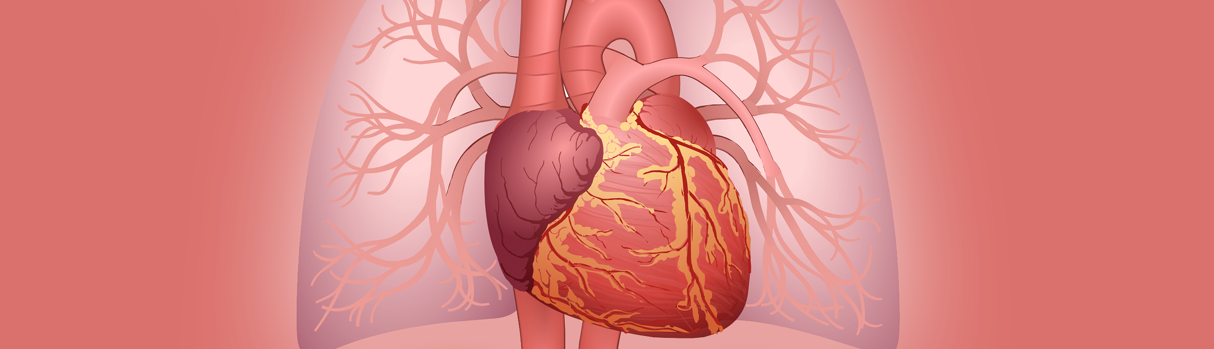
What are the parts of the heart?</h2> <figure class="asset-c-100"><span class="asset-image-title">Heart anatomy</span> <div class="asset-animation asset-cv-animation"> <iframe src="https://www.aboutkidshealth.ca/Style%20Library/akh/animation/HeartAnatomy%20-%20Storyline%20output/story.aspx"></iframe> </div></figure> <p>The right and left sides of the heart are divided by a wall called the septum. The right side pumps blood to the lungs, where the blood picks up oxygen. The left side pumps blood to the rest of the body.</p><p>The heart is made up of four hollow chambers:</p><ul><li>The upper two chambers are the right atrium and left atrium. These are called "collecting chambers" because they collect the blood as it returns to the heart.</li><li>The lower two chambers are the right ventricle and left ventricle. These are called "pumping chambers" because they pump the blood out of the heart to where it needs to go.</li></ul><p>Blood flows from chamber to chamber through the following valves, which keep blood flowing forward and prevent it from leaking backward: </p><ul><li>The tricuspid valve lets blood flow from the right atrium to the right ventricle.</li><li>The pulmonary valve lets blood flow from the right ventricle to the pulmonary artery.</li><li>The mitral valve lets blood flow from the left atrium to the left ventricle.</li><li>The aortic valve lets blood flow from the left ventricle to the aorta.</li></ul><h3>How does the heart pump blood?</h3> <figure class="asset-c-80"> <span class="asset-image-title">Normal heart anatomy</span><img src="https://assets.aboutkidshealth.ca/AKHAssets/Hearts_CHD/Normal_Heart_CHD.jpg" alt="Normal heart showing placement of the atria, ventricles, aorta, superior vena cava, inferior vena cava, pulmonary artery and pulmonary veins" /><figcaption class="asset-image-caption">The heart has two upper (receiving) and two lower (pumping) chambers. Blood flows into the upper chambers (the right atrium and the left atrium). The lower chambers (the right and left ventricles) pump blood out of the heart. The heart valves open and close to keep blood flowing in the correct direction. </figcaption> </figure> <p>After blood has travelled through the body, it comes back to the heart through the veins (vena cava) and enters the right atrium. The blood is bluish in colour because it is low in oxygen. When the heart relaxes, the blood flows through the tricuspid valve into the right ventricle.</p><p>The right ventricle contracts (squeezes) to send the blood through the pulmonary valve into the arteries in the lungs (pulmonary arteries). The blood travels to the lungs, where it gets fresh oxygen and turns bright red again. It returns to the left atrium through the pulmonary veins. Then it moves through the mitral valve to the left ventricle. The left ventricle pumps the blood high in oxygen out through the aortic valve into the aorta. The aorta delivers the blood to the body and the process begins again.</p><h3>What are the coronary arteries?</h3><p>The heart itself is a muscle, and it needs oxygen to work. The coronary arteries branch off from the aorta. They are the vessels that bring blood high in oxygen to the heart.</p><p>The main arteries are the right coronary artery and the left coronary artery.</p><ul><li>The right coronary artery brings blood to the ventricles, right atrium, and sinoatrial node.</li><li>The left coronary artery branches into the circumflex artery and the left anterior descending artery. The left coronary artery supplies the blood to the ventricles and left atrium.</li></ul><p>This blood flow is known as coronary circulation.</p><h2>What is a heartbeat?</h2><p>As the heart beats, it makes a “lub-dub” sound. This is the sound made by the heart valves as they open and close. With each heartbeat, blood pushes through the aortic valve into the aorta and is delivered to the body. The heart beats about 100,000 times a day.</p><h3>What is a pulse?</h3><p>You can feel a pulse, the rhythmic beating of the heart, each time the ventricle contracts by touching your index and middle finger (not your thumb) to your wrist or the side of your neck. To figure out what your heart rate is, keep your fingers on your pulse and count the beats for 10 seconds, then multiply by 6.</p><ul><li>A newborn's heart rate is about 130 to 160 beats per minute (BPM) at rest</li><li>A 6-month-old's heart rate is about 100 BPM</li><li>A toddler's heart rate is about 70 to 80 BPM</li><li>An adult's heart rate is about 60-80 BPM </li></ul><h2>How does the heart's electrical system work?</h2> <figure class="asset-c-80"> <span class="asset-image-title">The heart's electrical system</span><img src="https://assets.aboutkidshealth.ca/AKHAssets/Hearts_EP/EP_heart_Normal.jpg" alt="Normal heart showing placement of the SA node, AV node, His bundle and Purkinje fibers" /><figcaption class="asset-image-caption">1) An electrical signal starts in the sinoatrial (SA) node, which signals the atria to contract. 2) The electrical signal moves from the SA node to the atrioventricular (AV) node. 3) From the AV node, the signal travels to the His bundle and then to the Purkinje fibers. 4) As this electrical signal moves through the heart, it stimulates the heart muscle to beat in a specific order; first the atria contract, followed by the ventricles. </figcaption> </figure> <p>There is a small area of the right atrium called the sinoatrial (SA) node. The SA node is the body’s natural pacemaker since it controls the heartbeat.</p><p>With each heartbeat, an electrical impulse from the SA node causes the muscles of the atria to contract. This lets them pump blood to the ventricles. The electrical signal is then carried through the atrioventricular (AV) node into the ventricles, causing them to contract and pump blood out of the heart. It's similar to the motion involved when you clench and unclench your fist.</p><p>Interfering with this electrical impulse can cause <a href="/article?contentid=890&language=english">problems with the heart rhythm (arrhythmias)</a> or stop the heartbeat altogether (cardiac arrest).</p><p>In cardiology, the area of practice related to heart rhythms and the heart’s electrical system is referred to as electrophysiology. </p><h2>What is blood pressure?</h2> <figure> <span class="asset-image-title">Blood pressure cuff</span><img alt="Child wearing blood pressure cuff" src="https://assets.aboutkidshealth.ca/AKHAssets/IMD_blood_pressure_cuff_EN.jpg" /> </figure> <p>Blood pressure is a measure of the pressure of the blood against the walls of the arteries. It is expressed as a top number and bottom number. Blood pressure indicates how well the heart is pumping.</p><p>To measure your child’s blood pressure, the nurse or doctor puts a blood pressure cuff on your child’s arm. The first number measured is the systolic pressure. This is the pressure as the heart pumps blood out. The second number is the diastolic pressure. This is the pressure as the heart relaxes and refills with blood. An average adult blood pressure is 120/80. That means 120 systolic and 80 diastolic.</p><p>Children's blood pressures vary with age and activities, such as exercise and sleep. Fever can also affect a child’s blood pressure. Some typical values are:</p><ul><li>75/50 for a newborn</li><li>96/65 for a toddler</li><li>100/60 for a preschooler</li><li>105/60 for a school-age child (6 to 8 years)</li><li>118/60 for a teenager</li></ul><p>Different factors can cause blood pressure to be too high or too low. Sometimes these factors are heart-related and sometimes they are not. Your child’s health-care provider can help you understand what an appropriate blood pressure value is for your child.</p>https://assets.aboutkidshealth.ca/akhassets/Hearts_CHD/Normal_Heart_CHD.jpgThe normal heartFalse

Thank you to our sponsors

AboutKidsHealth is proud to partner with the following sponsors as they support our mission to improve the health and wellbeing of children in Canada and around the world by making accessible health care information available via the internet.

Our Sponsors